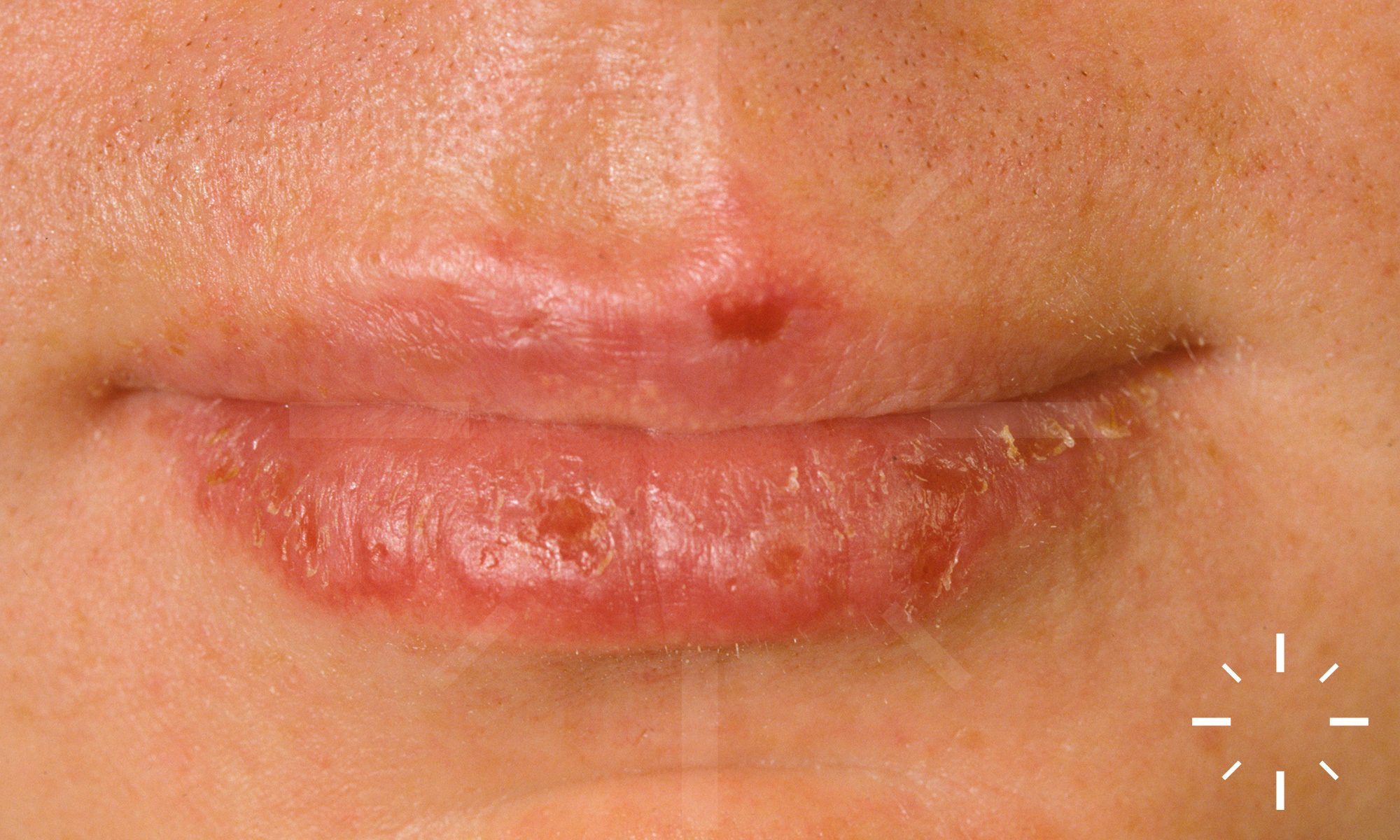
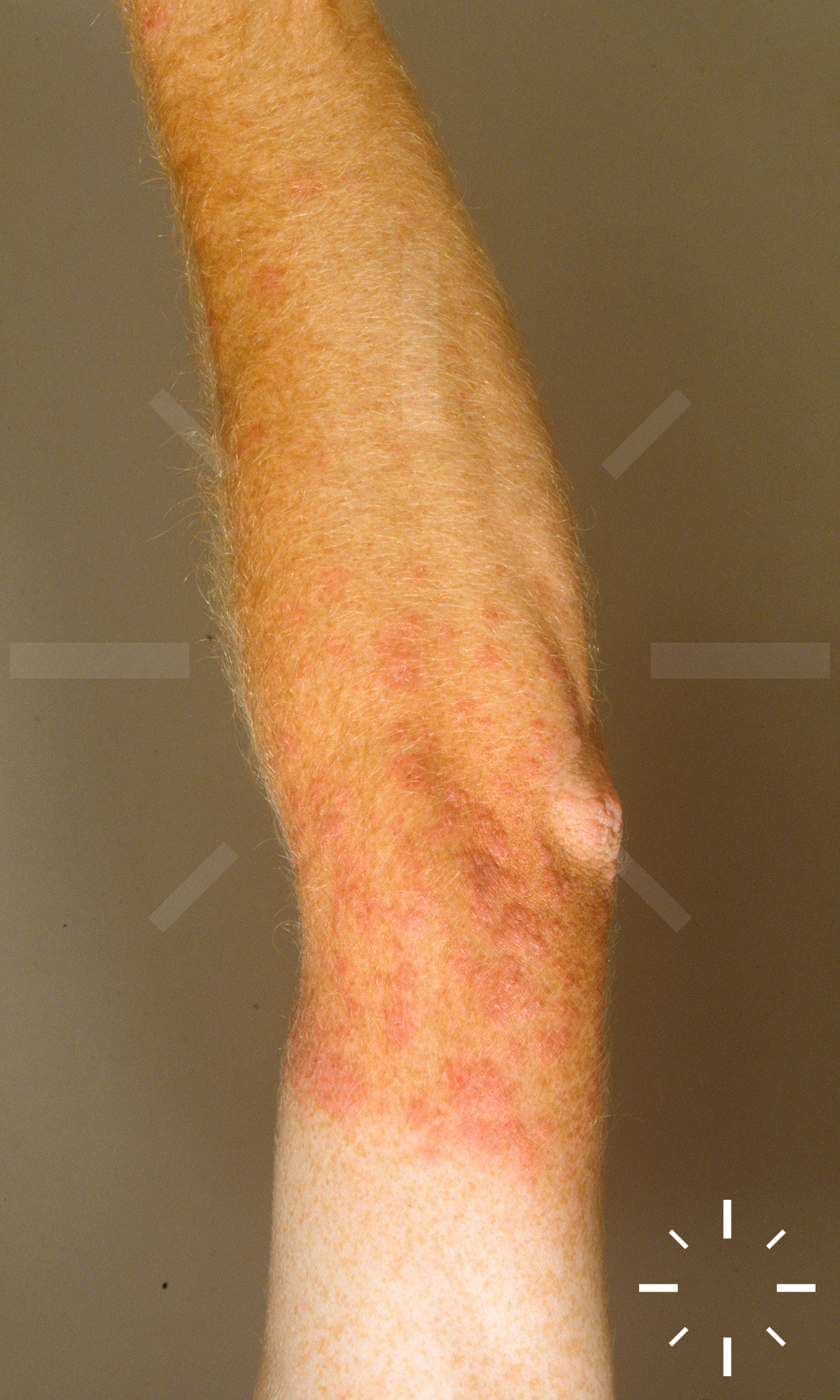
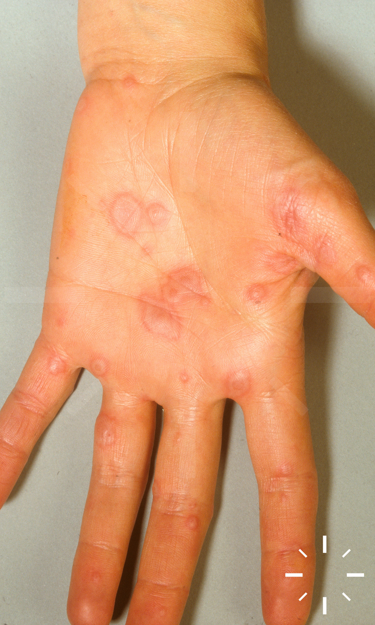
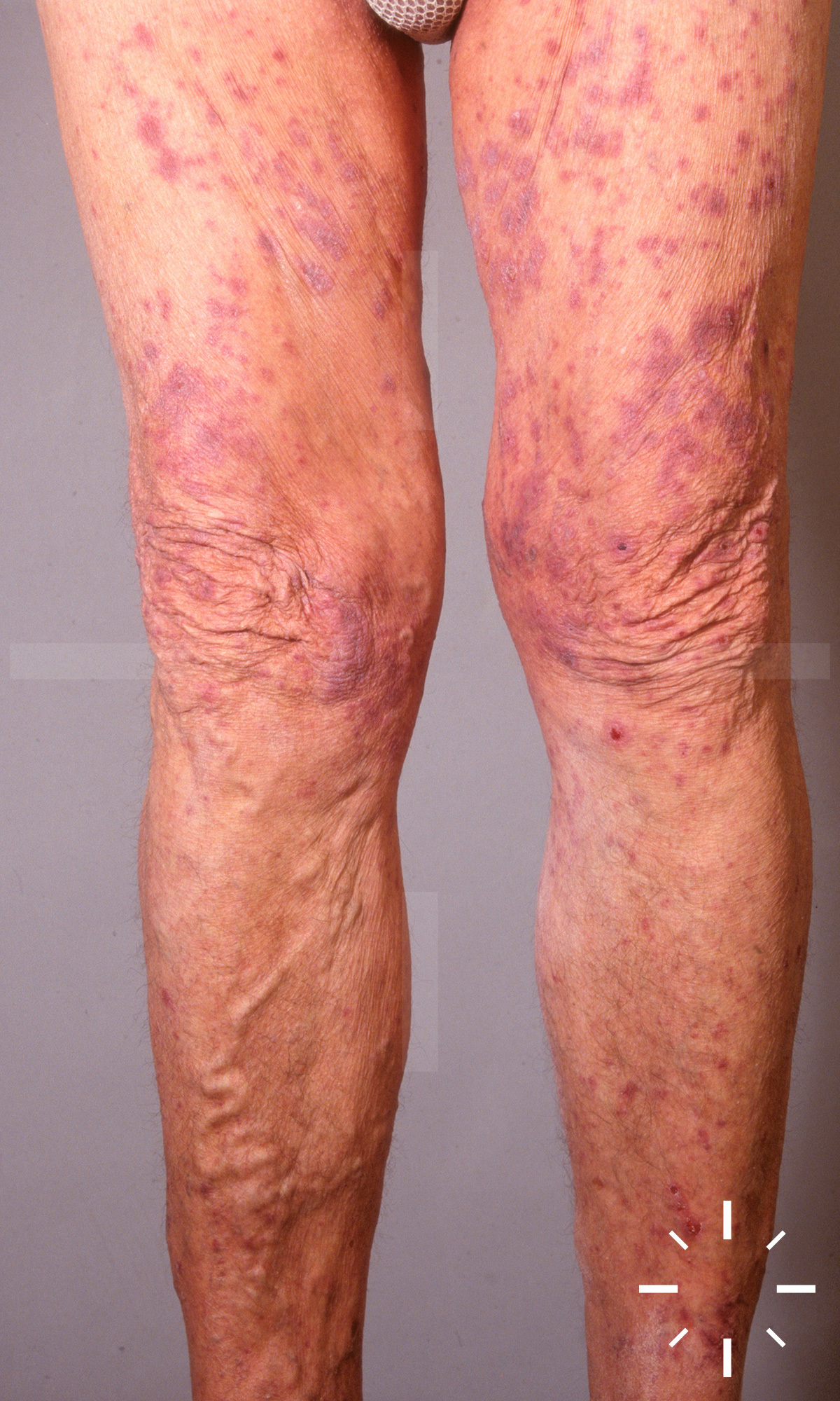
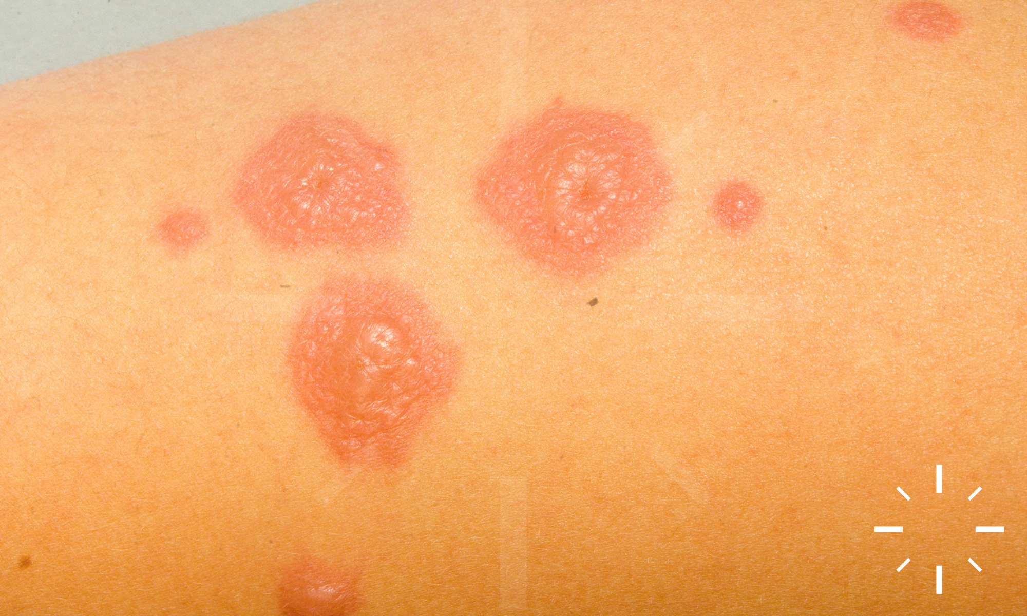
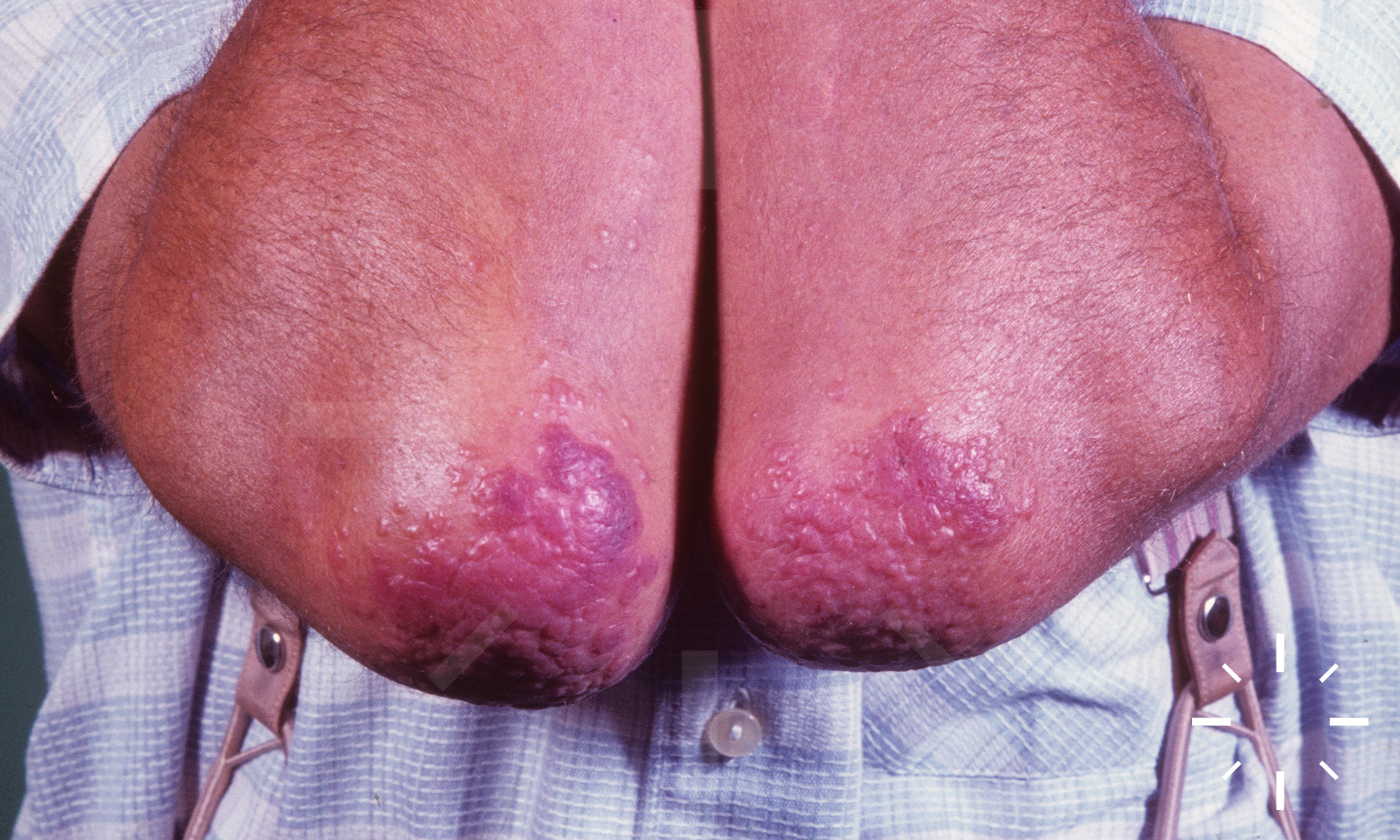
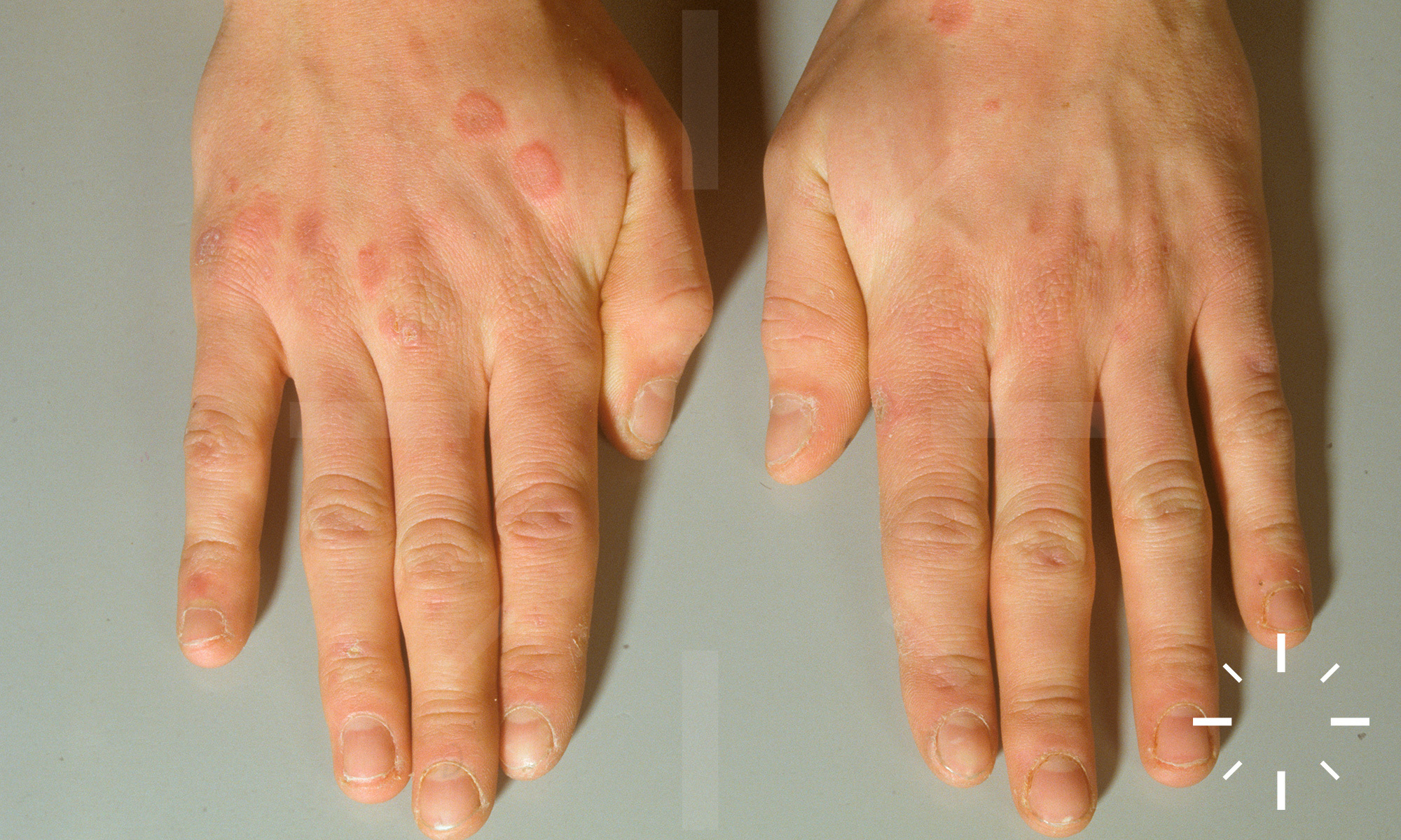
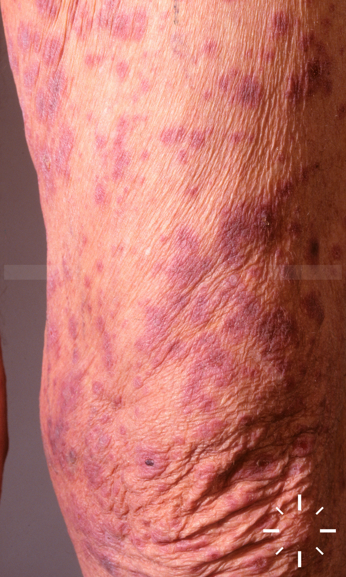
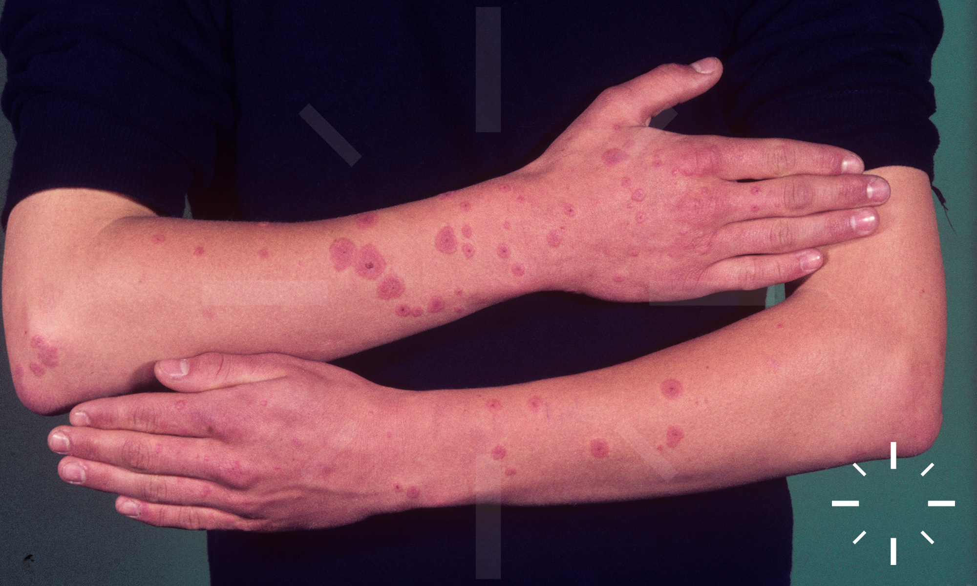
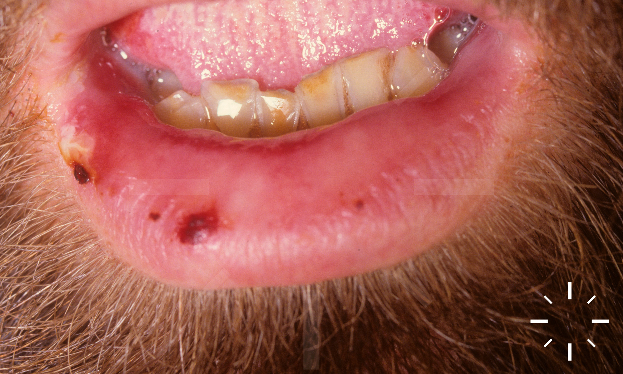
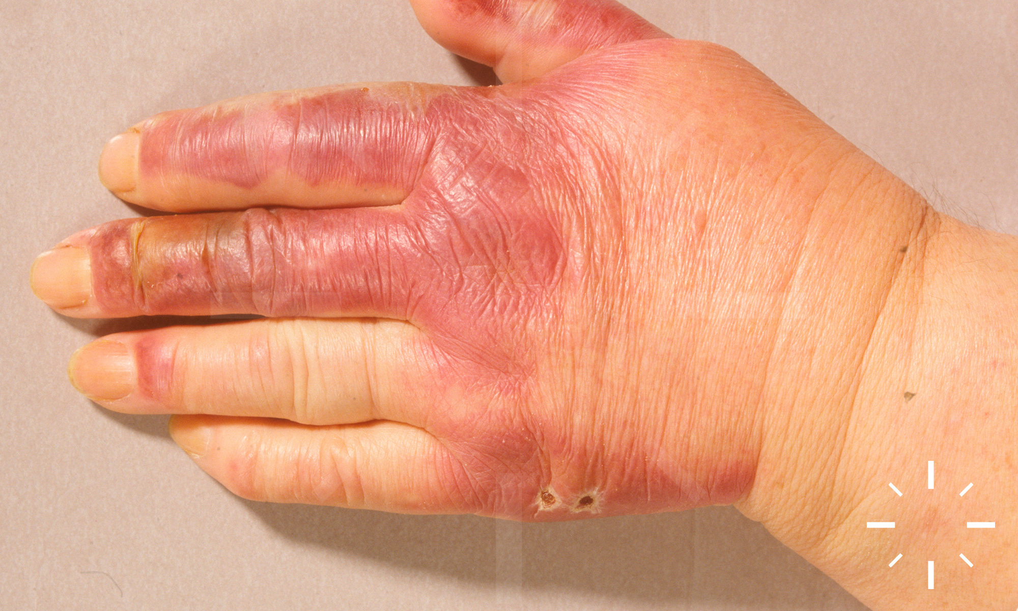
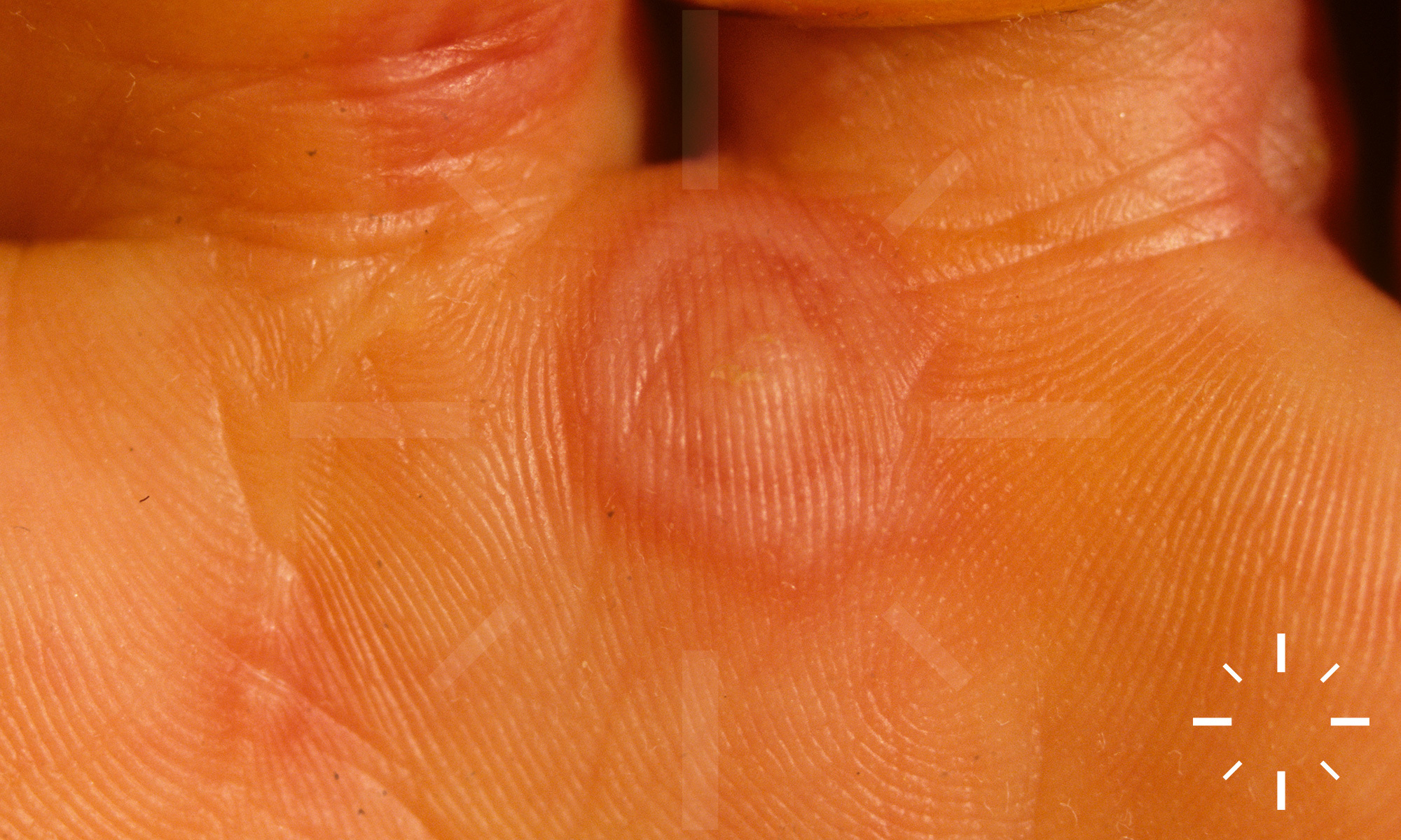
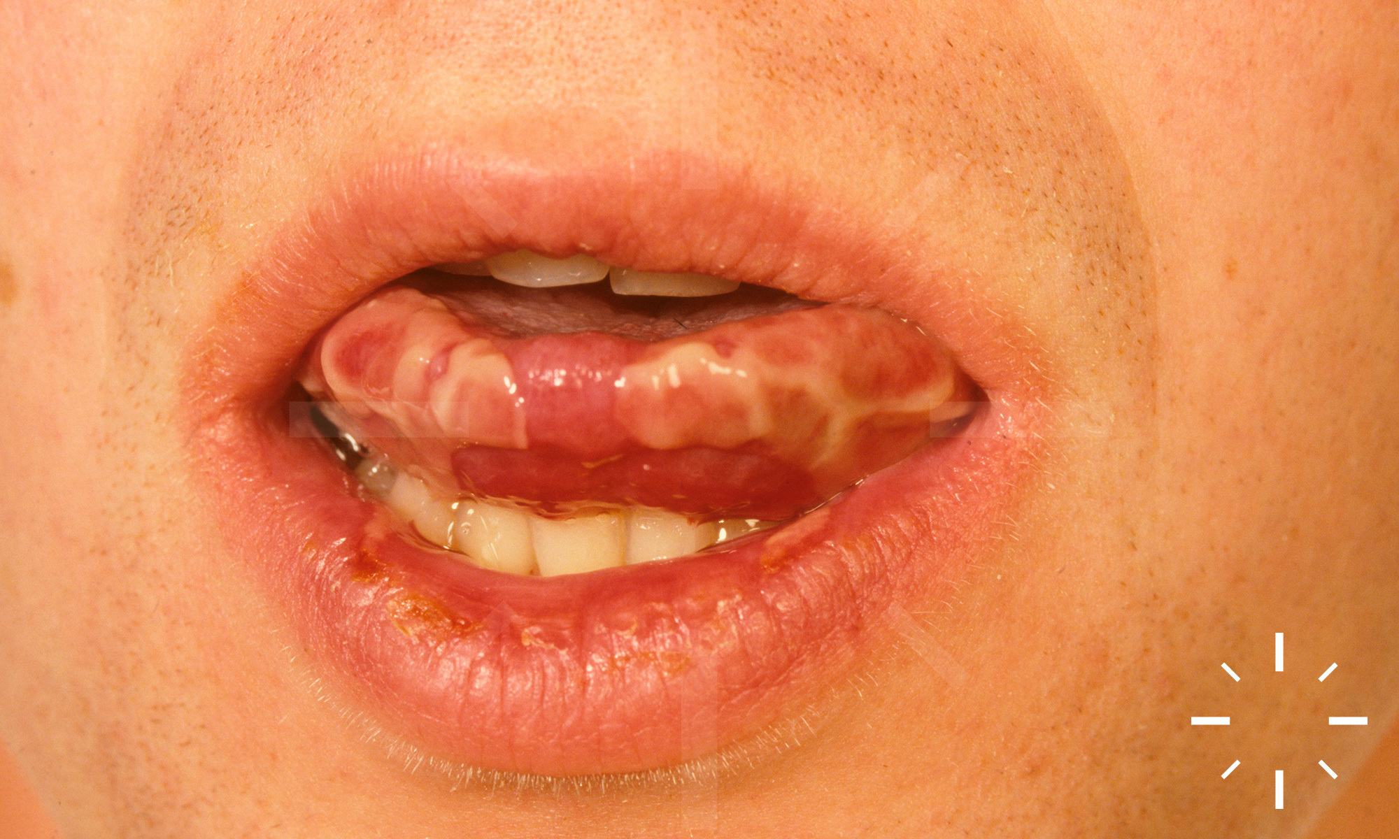
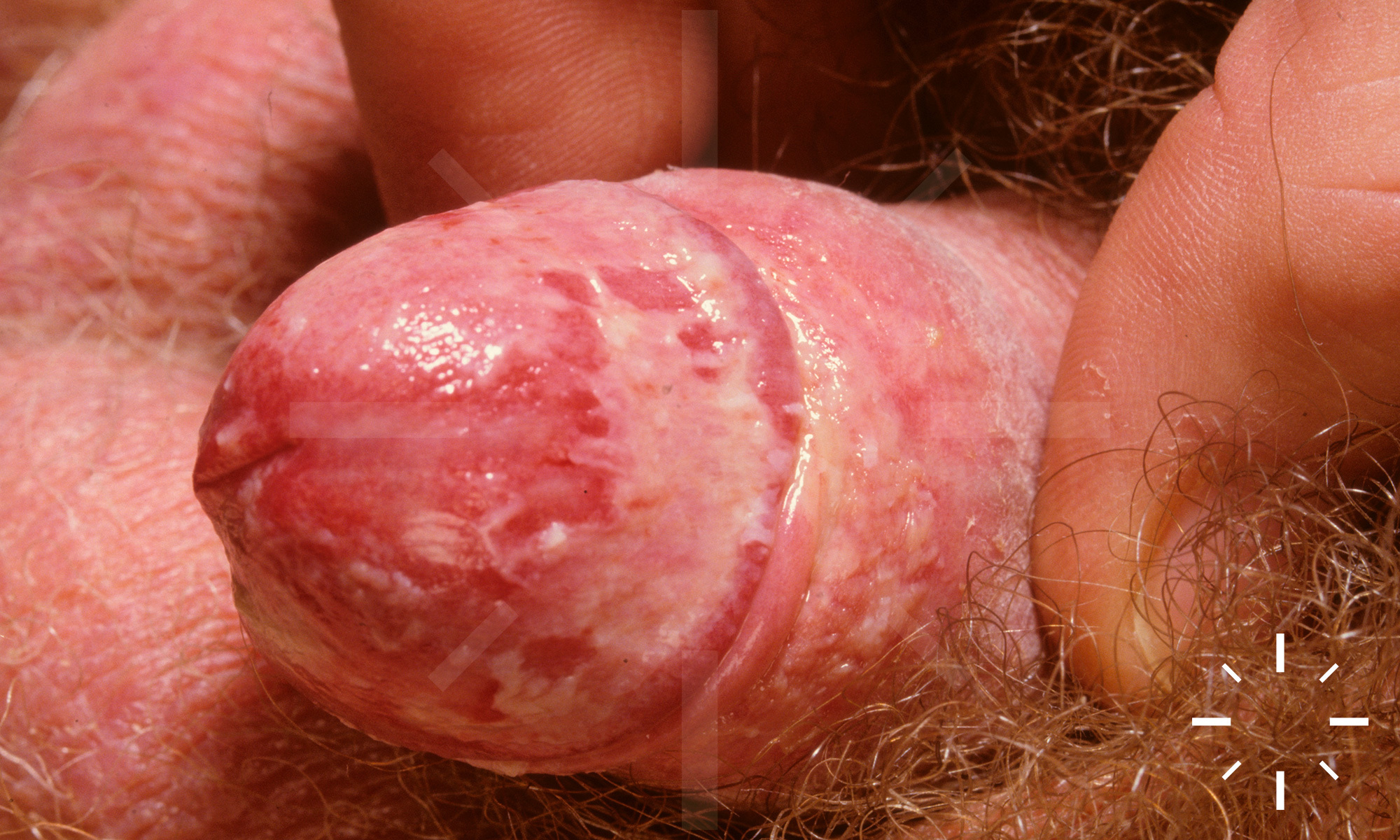
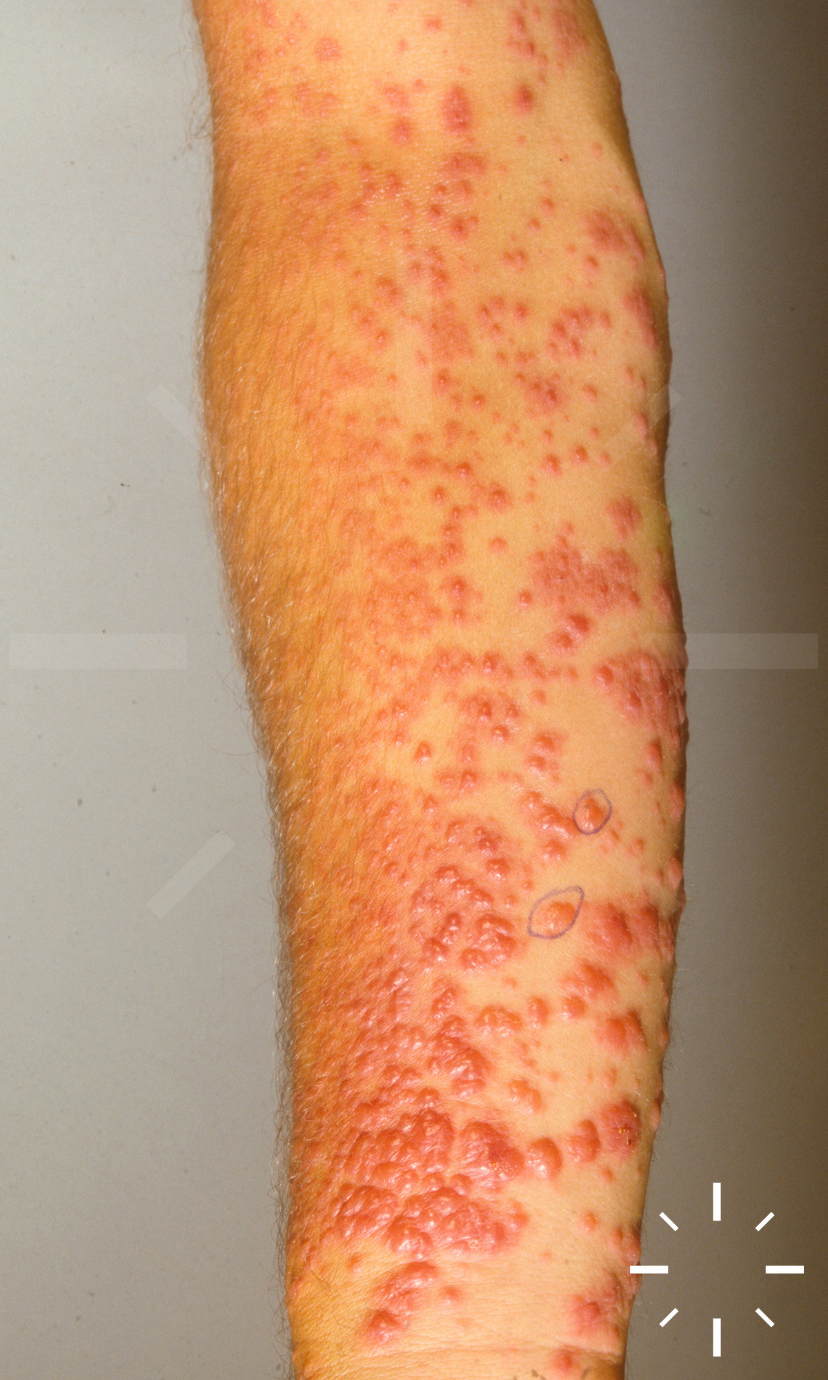
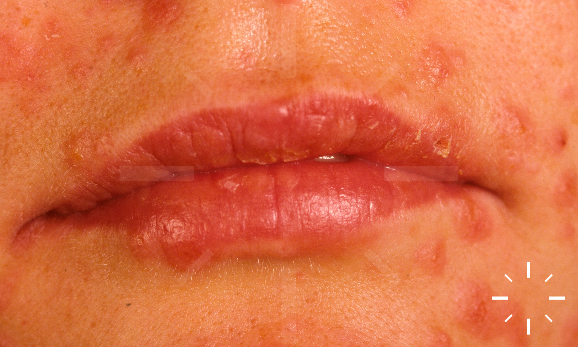
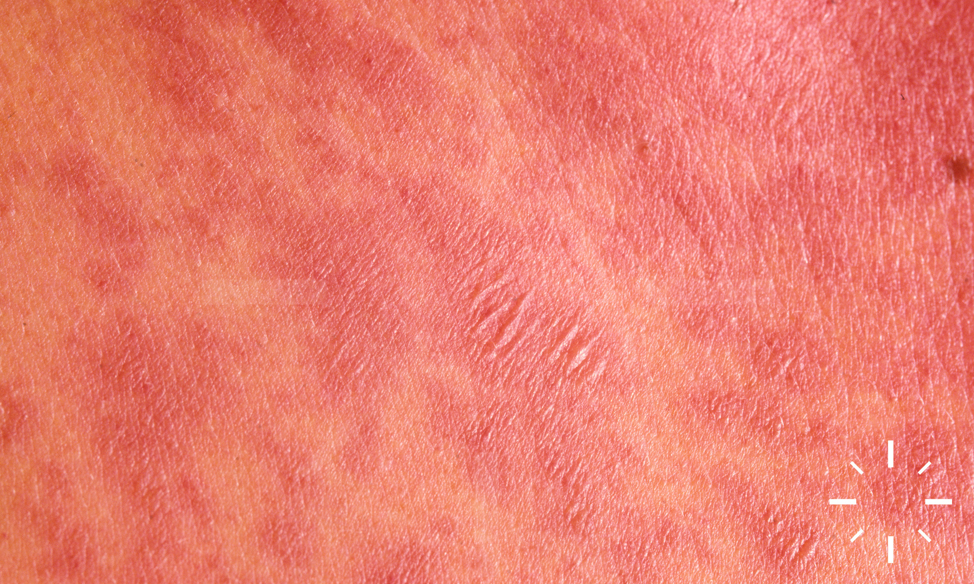
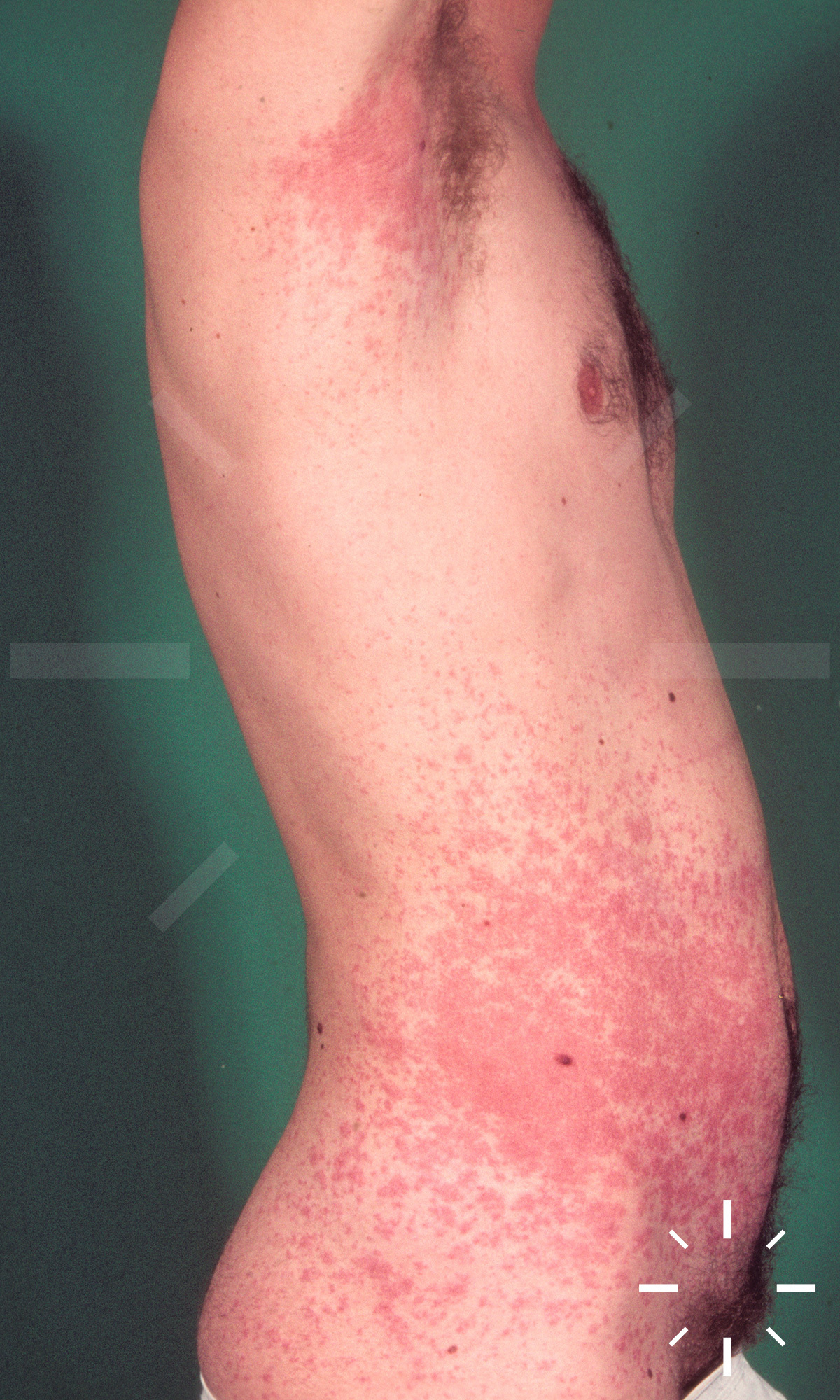
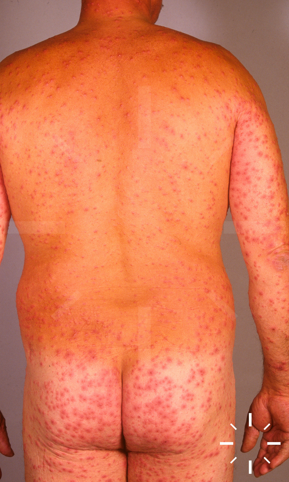
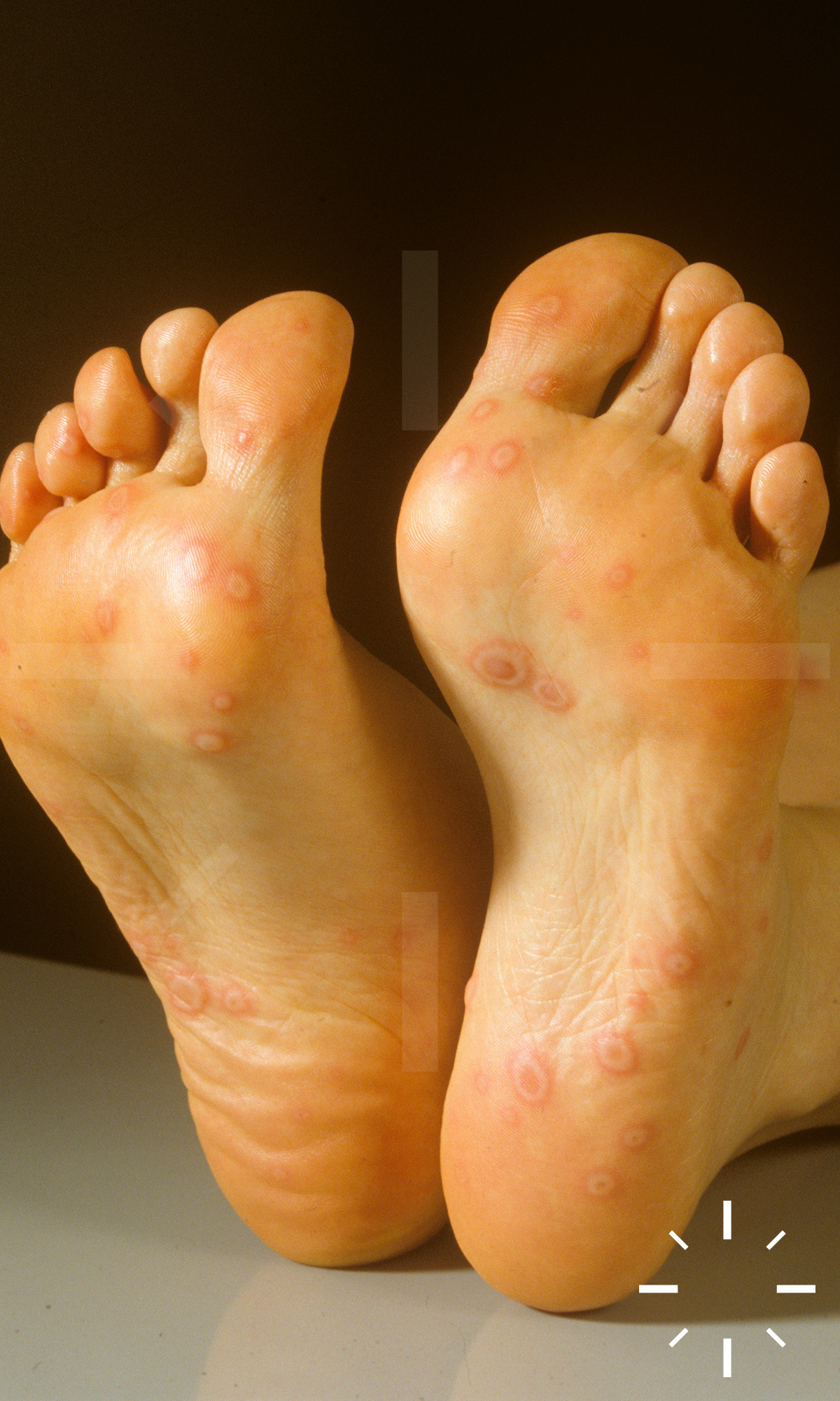
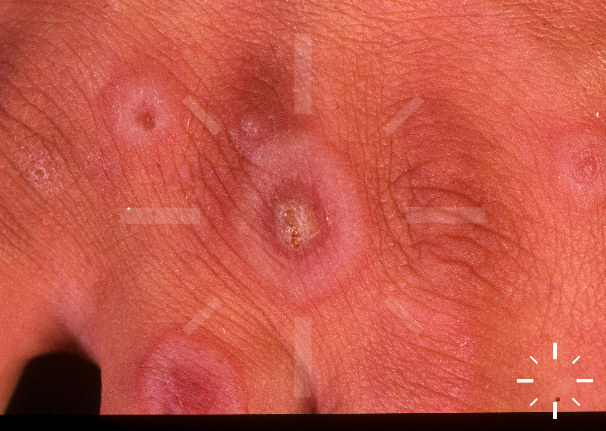
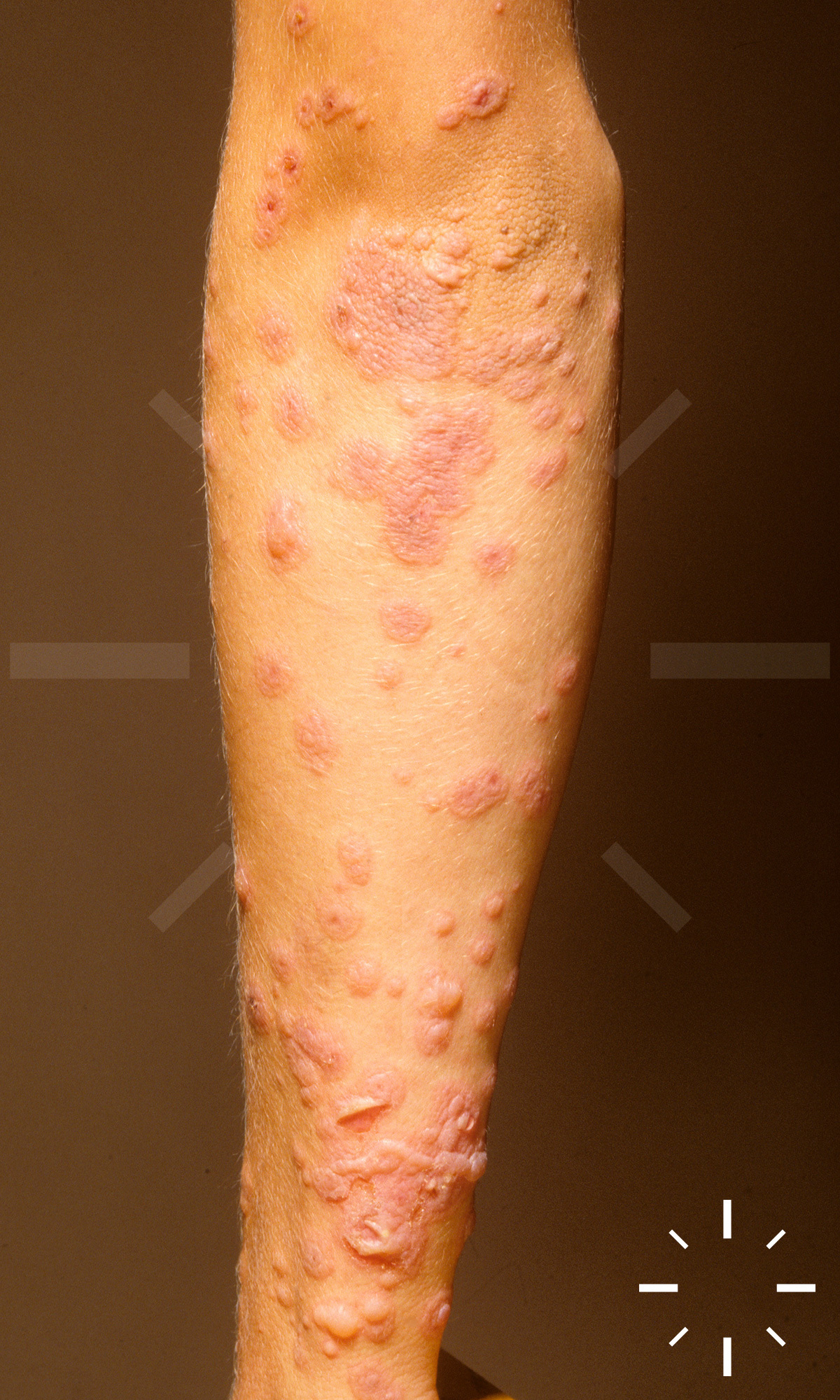
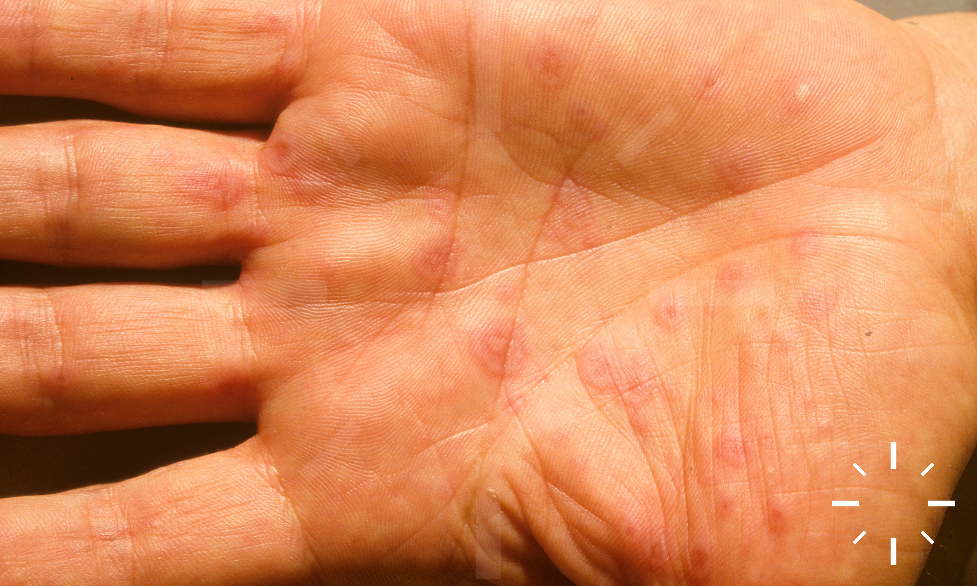
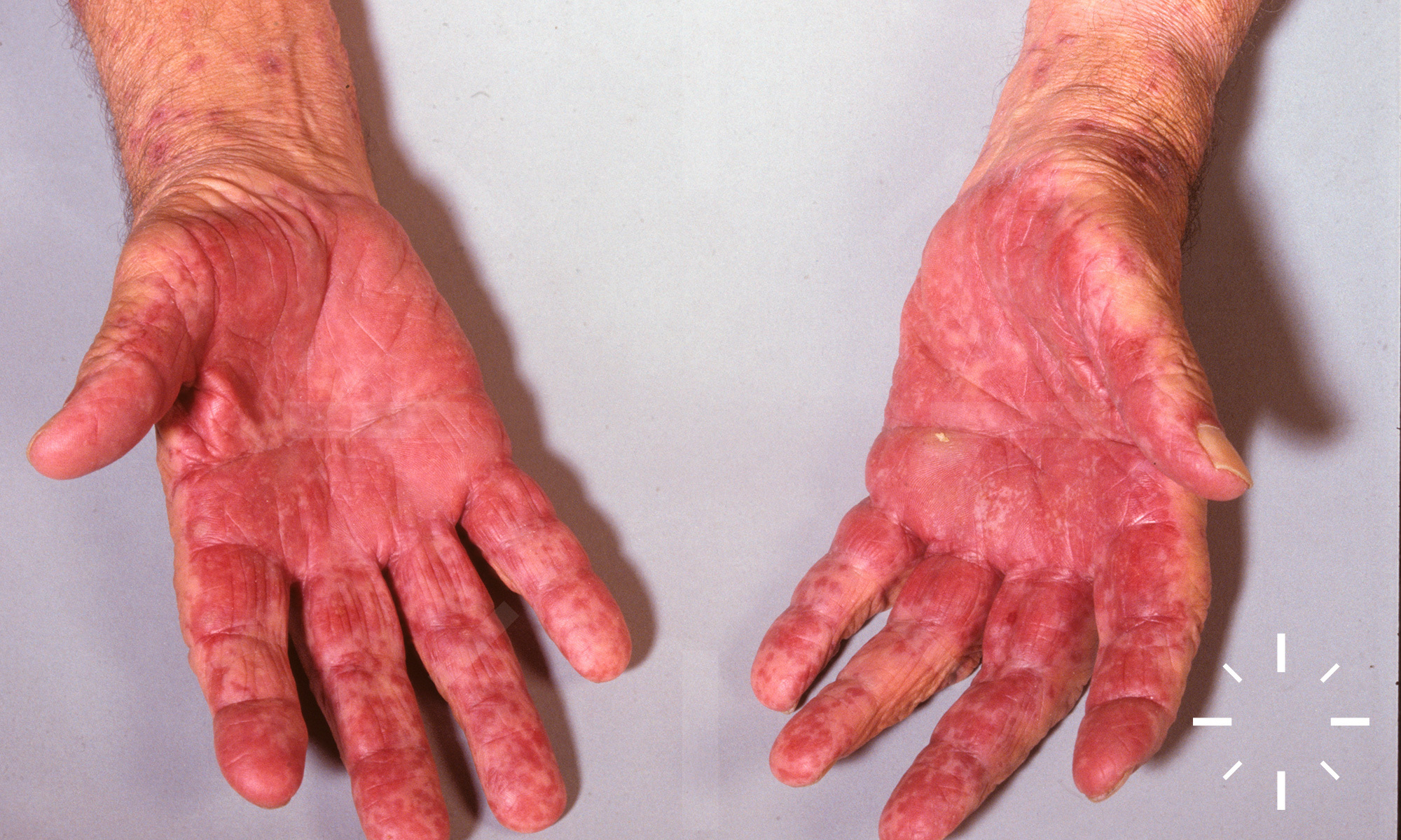
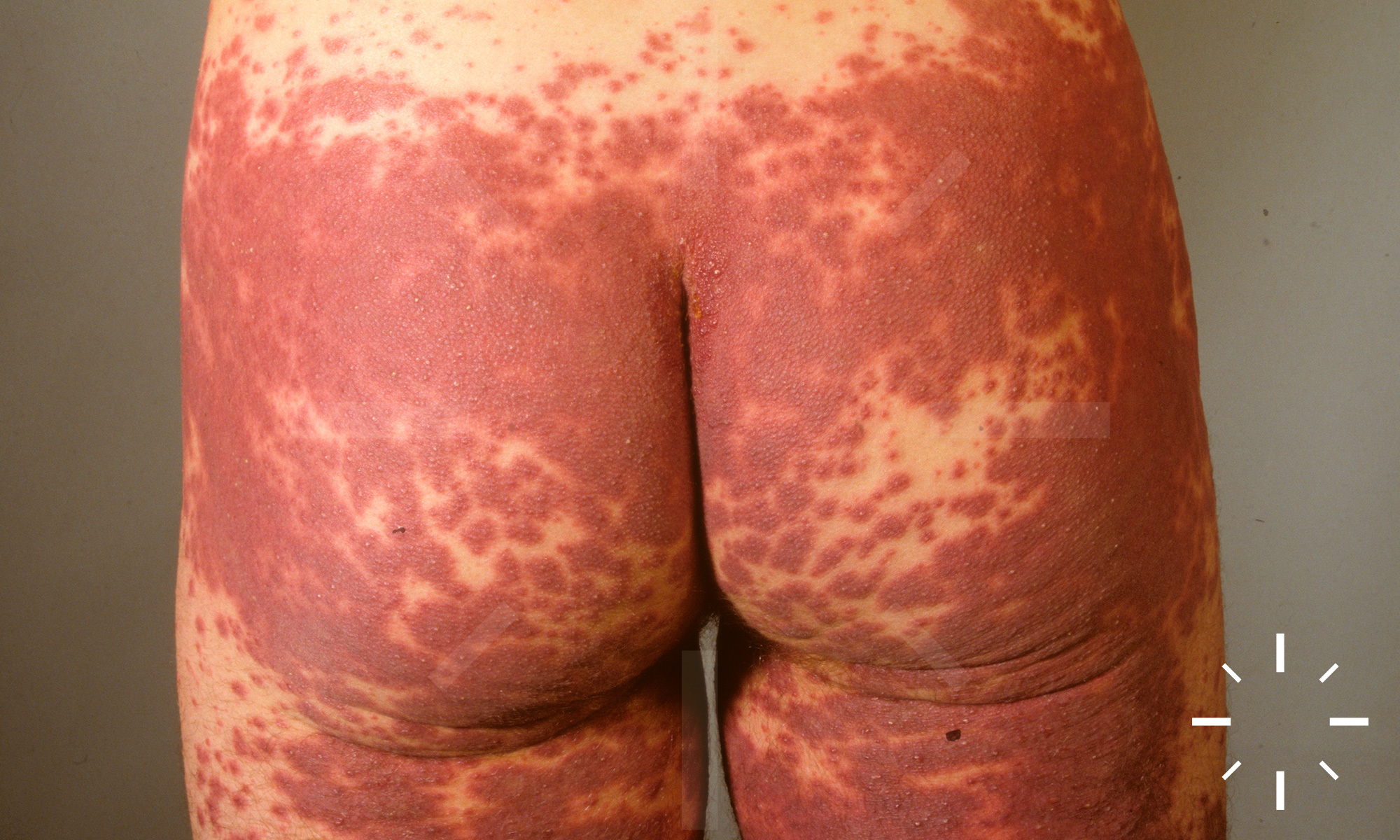
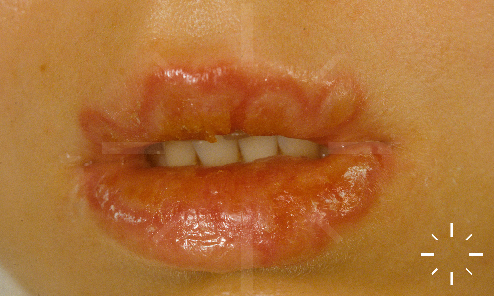
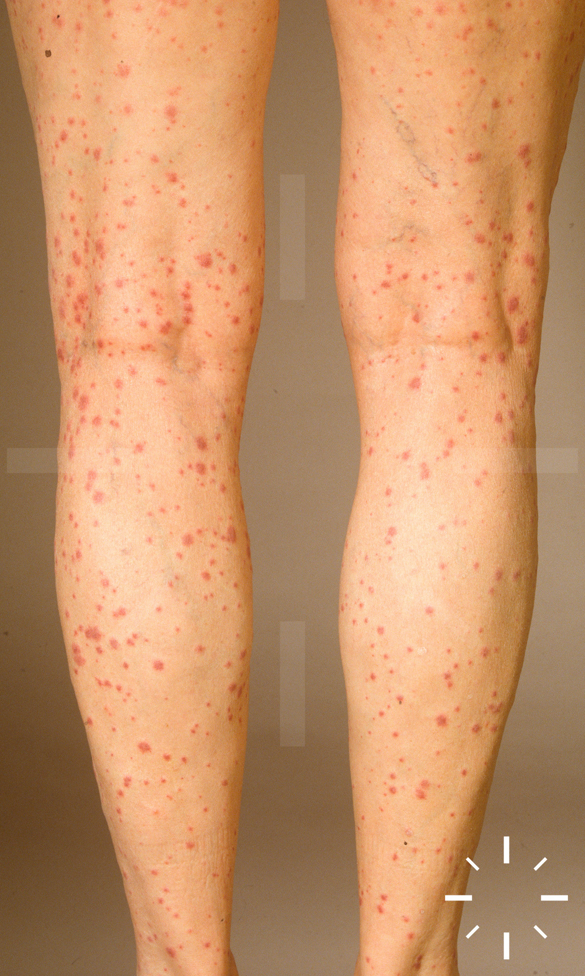
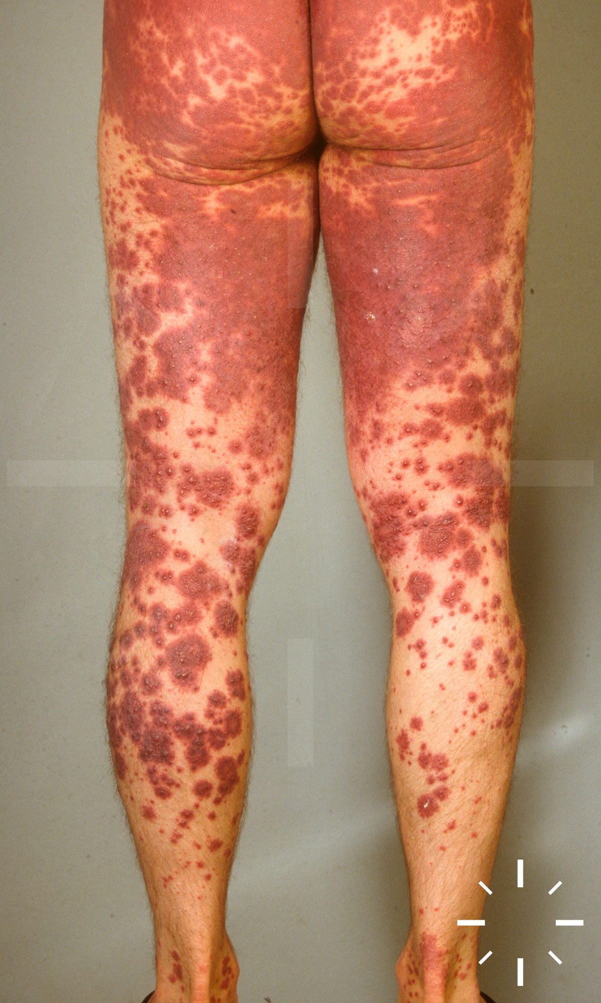
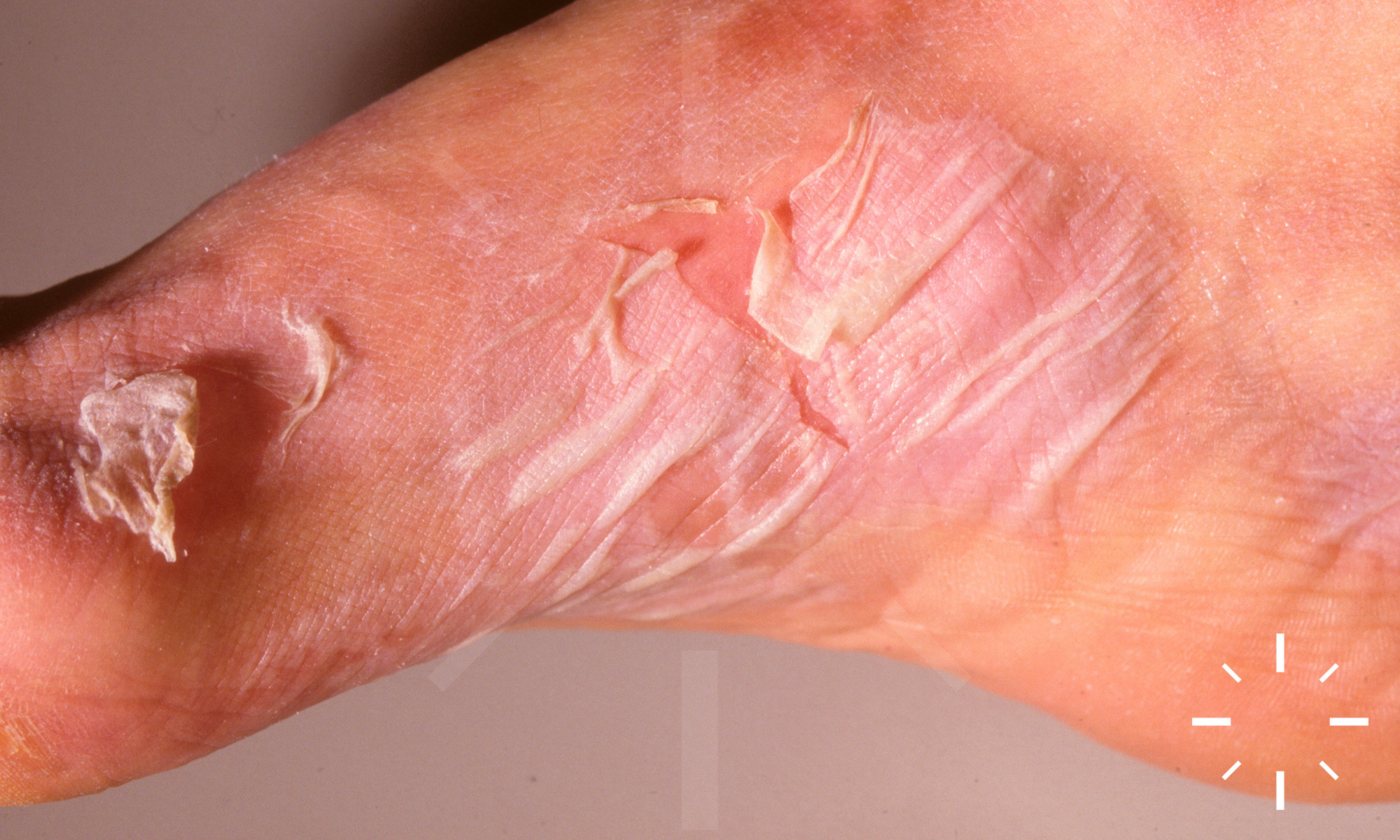
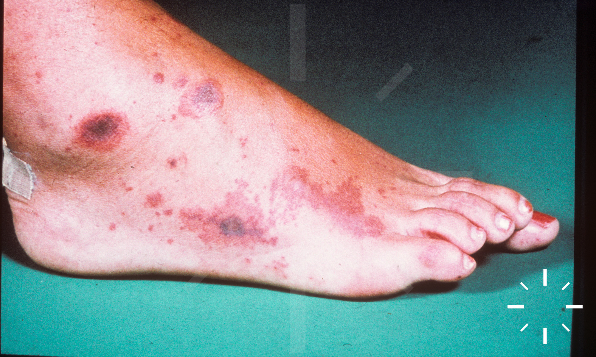
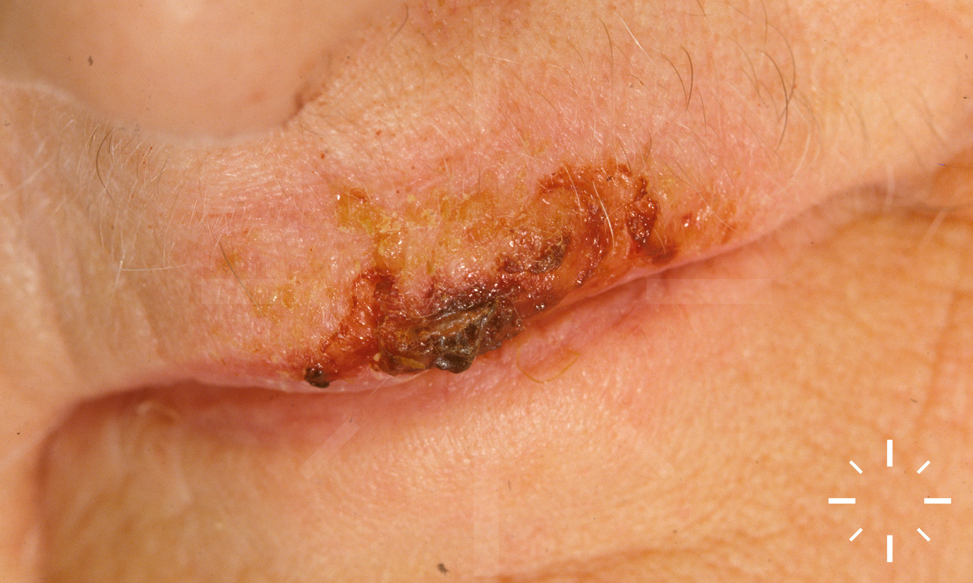
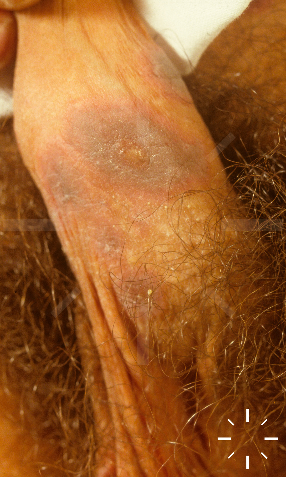
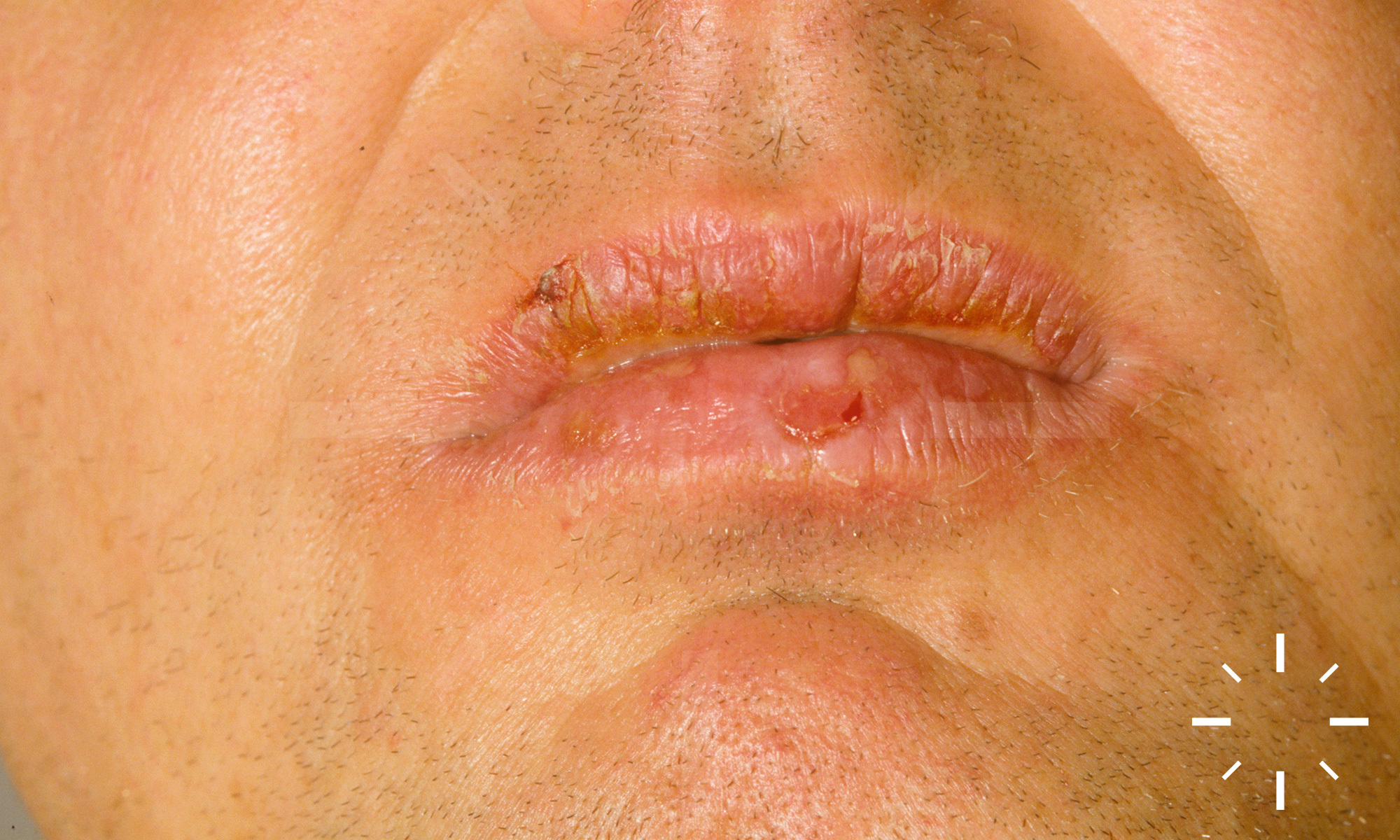
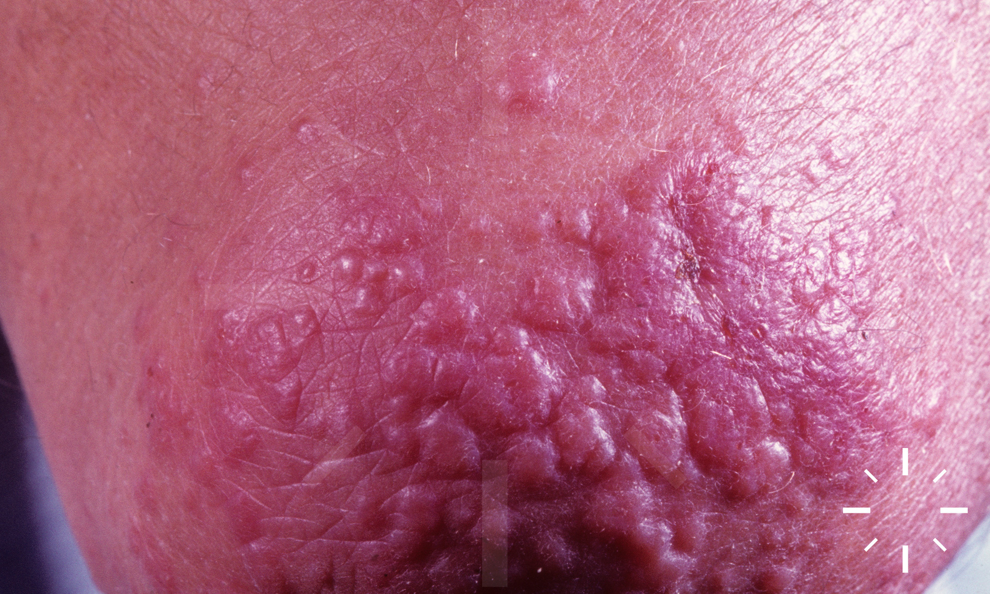
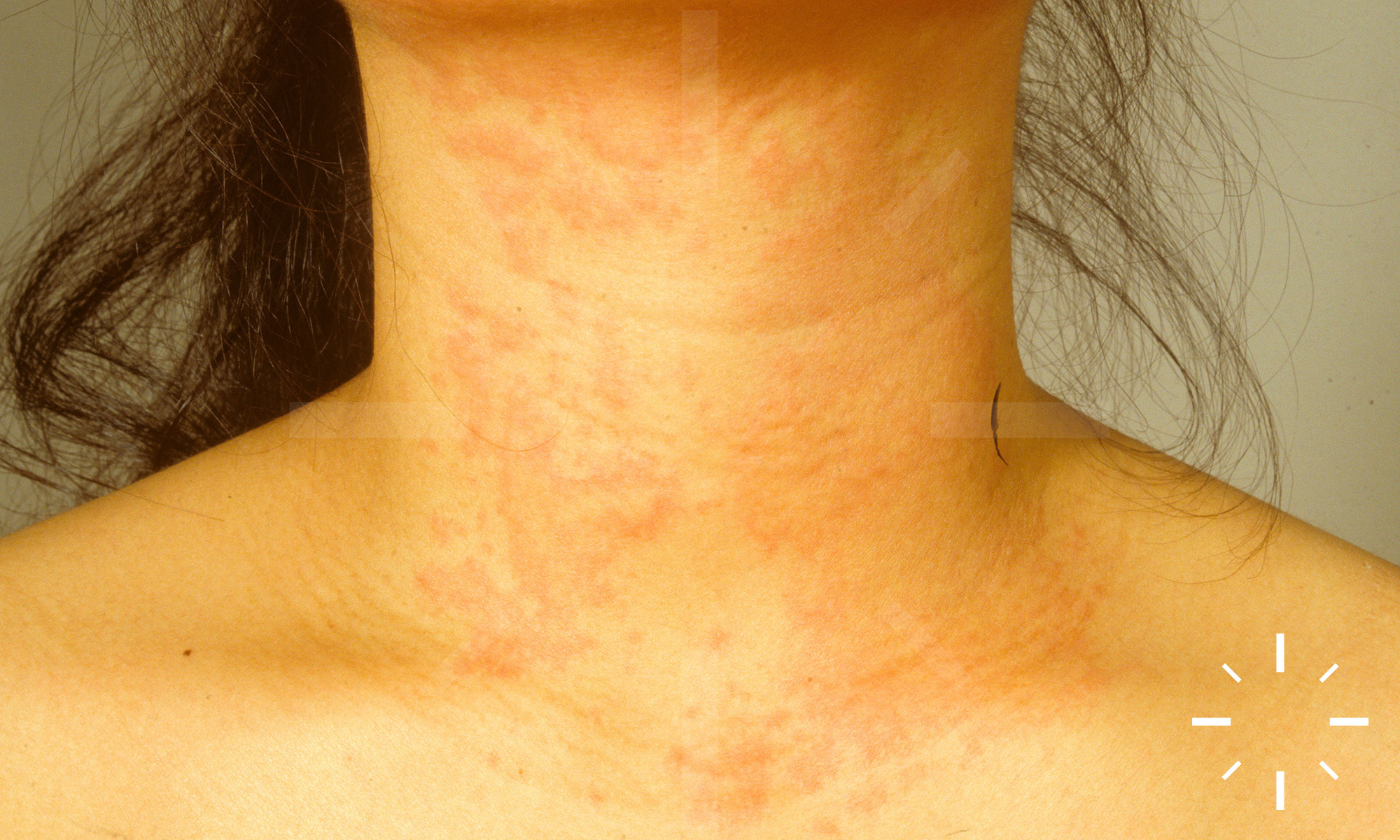
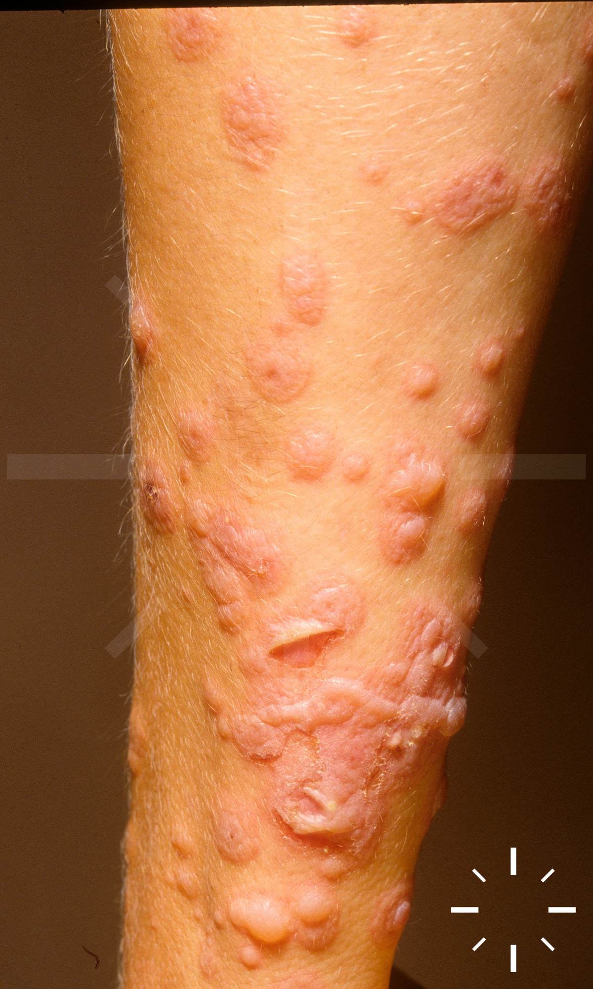
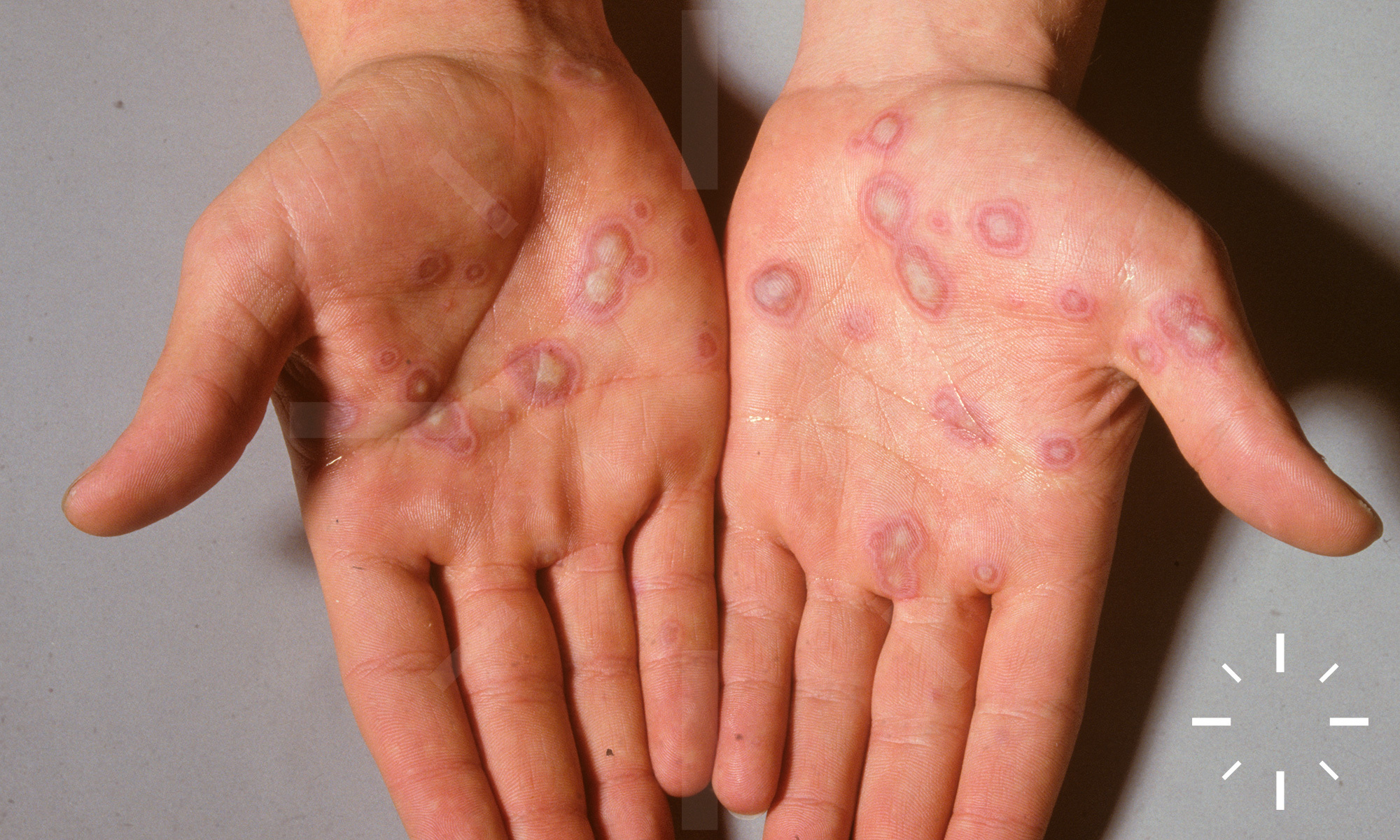
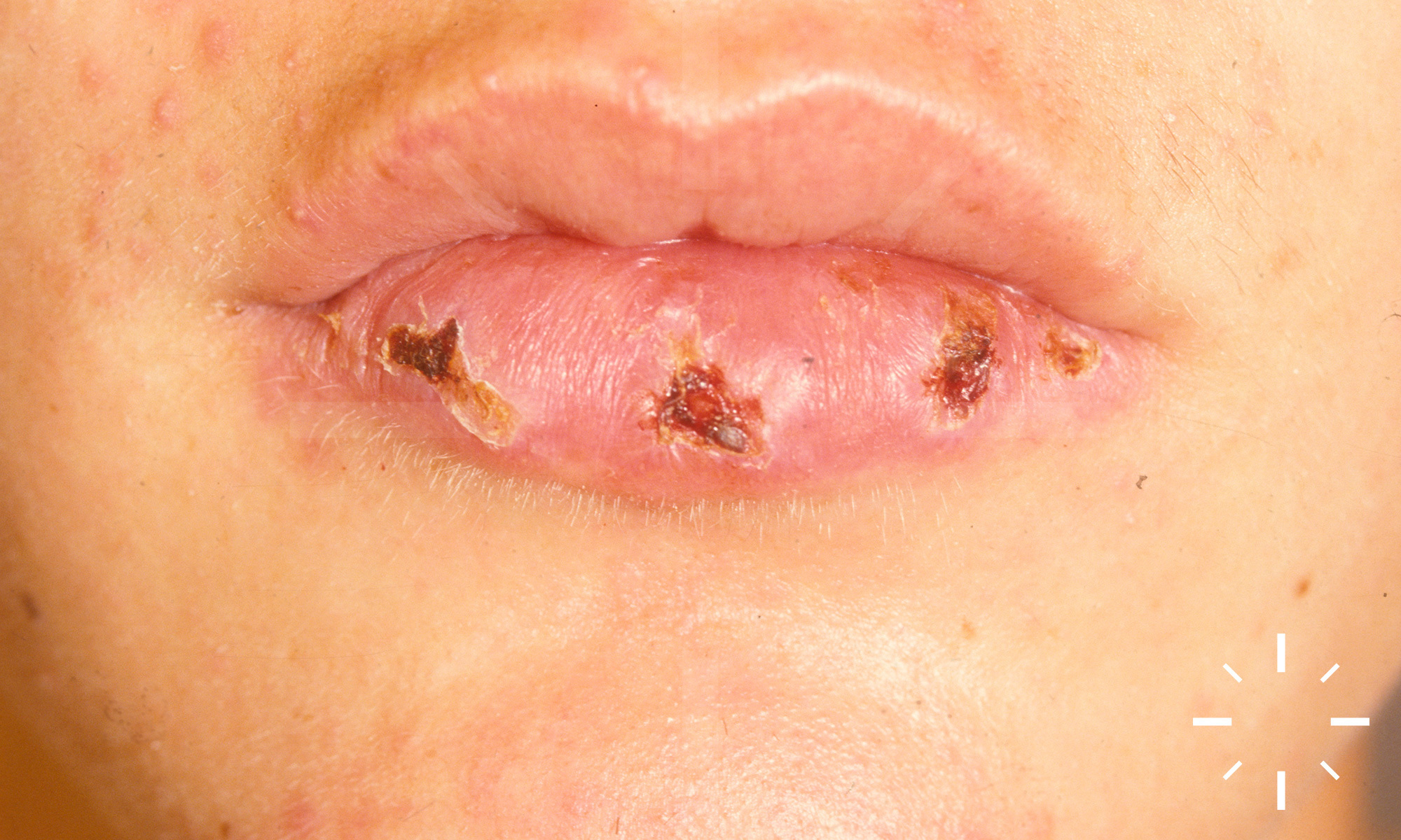
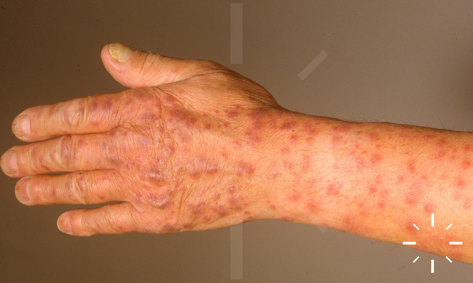
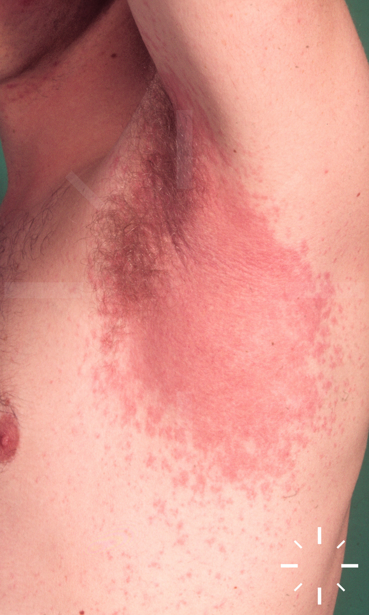
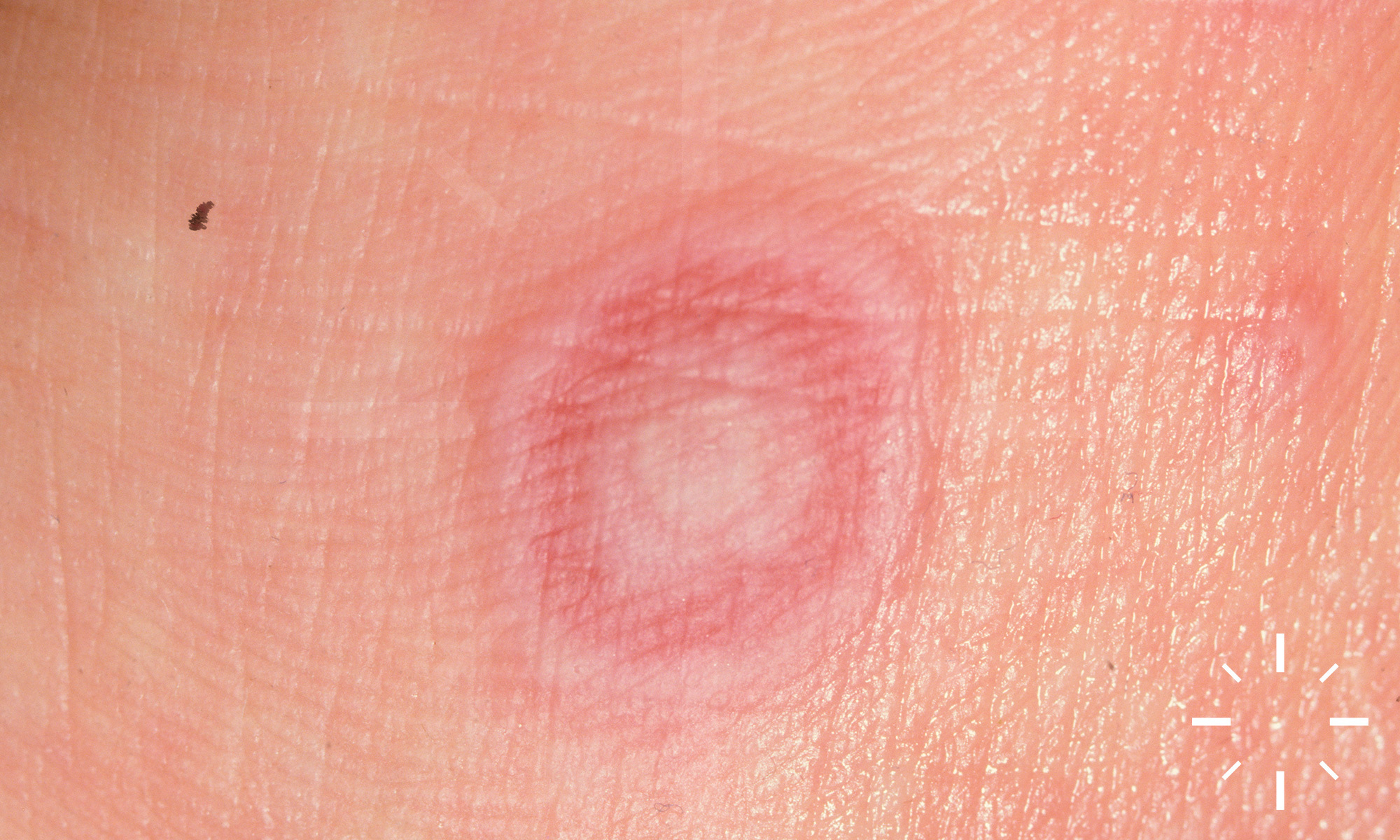
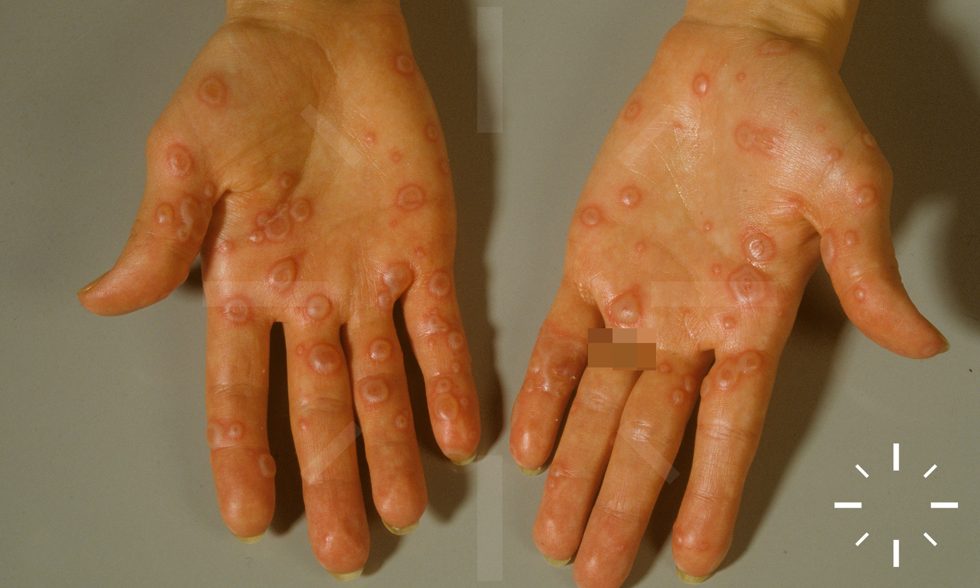
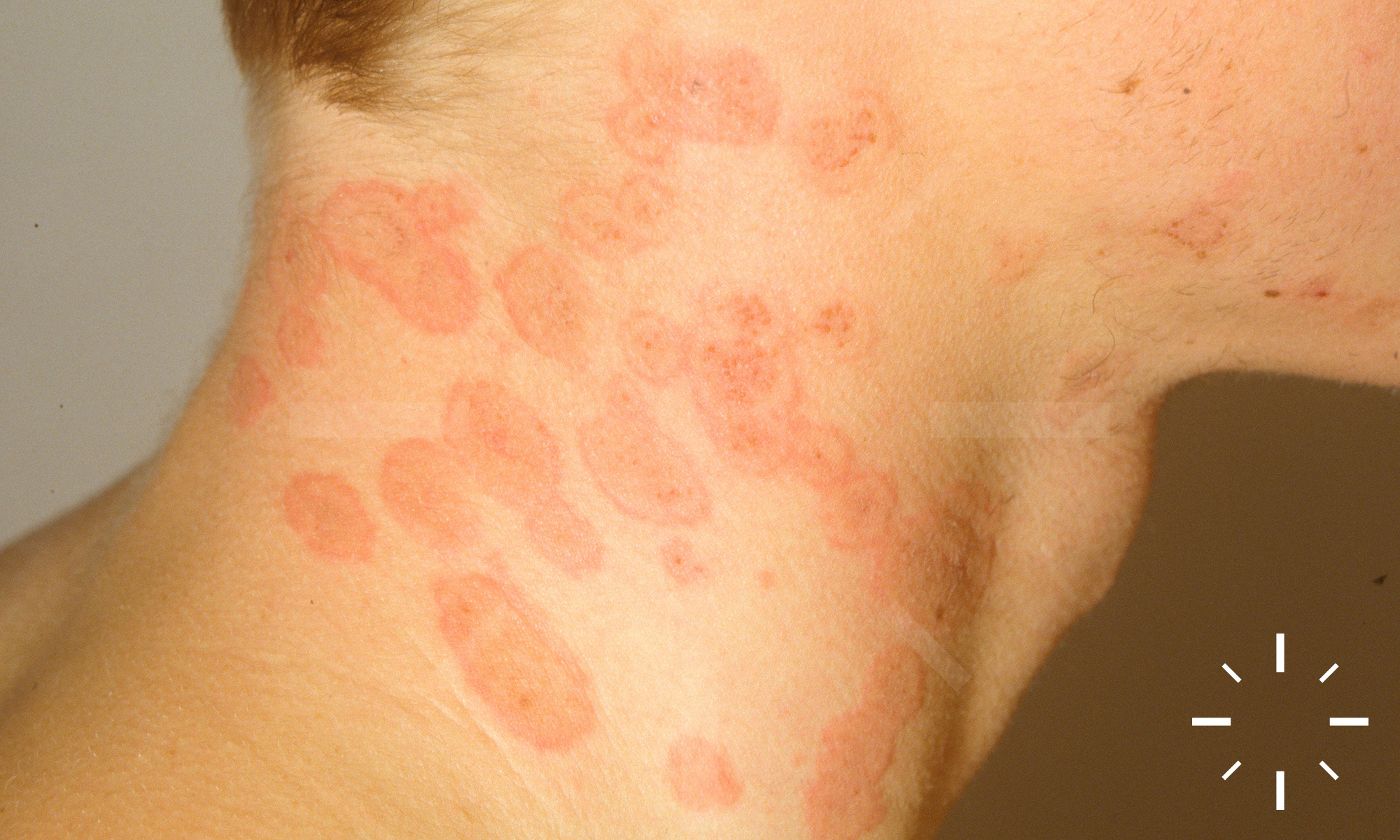
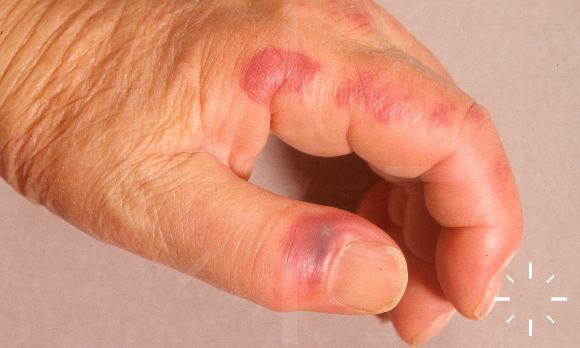
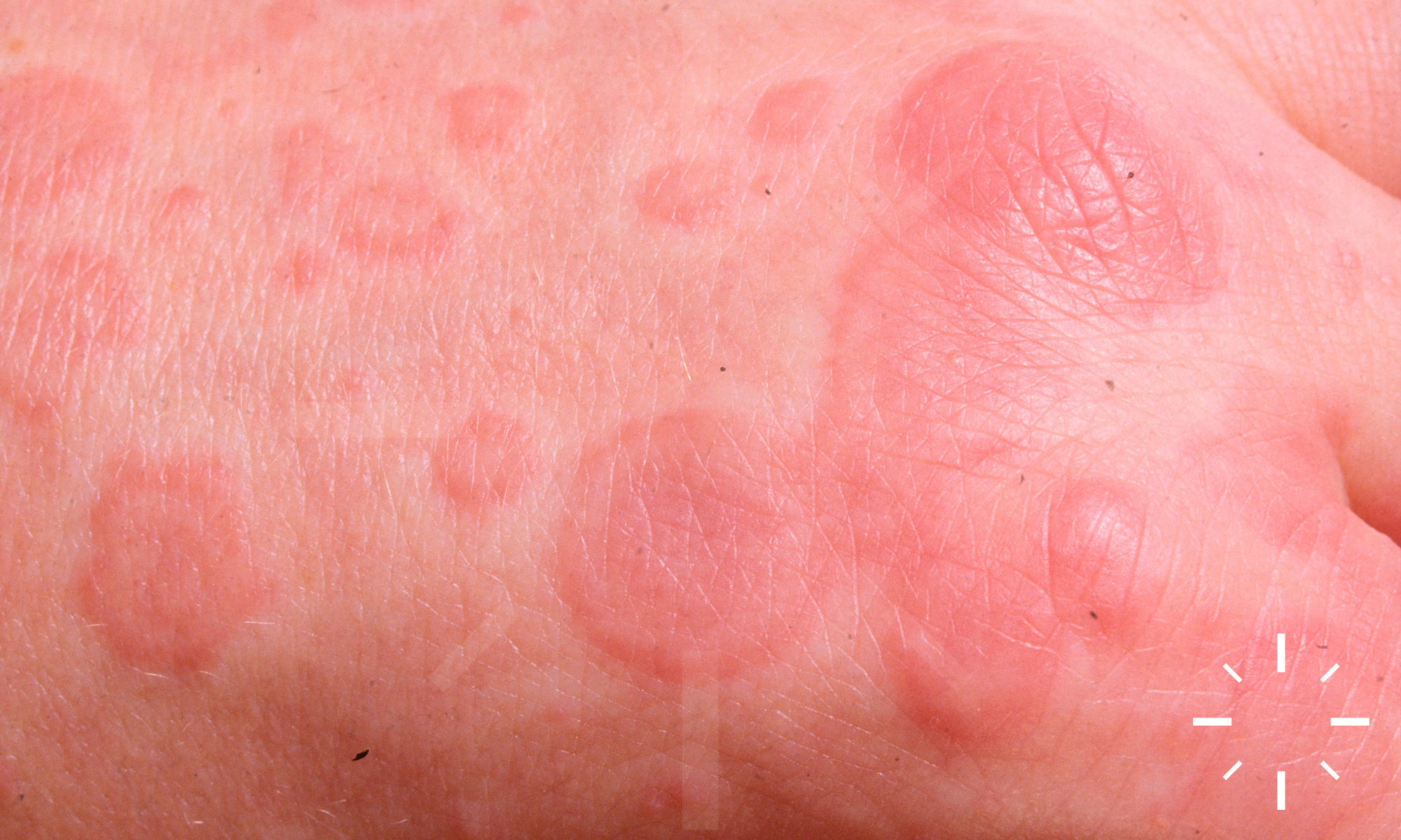
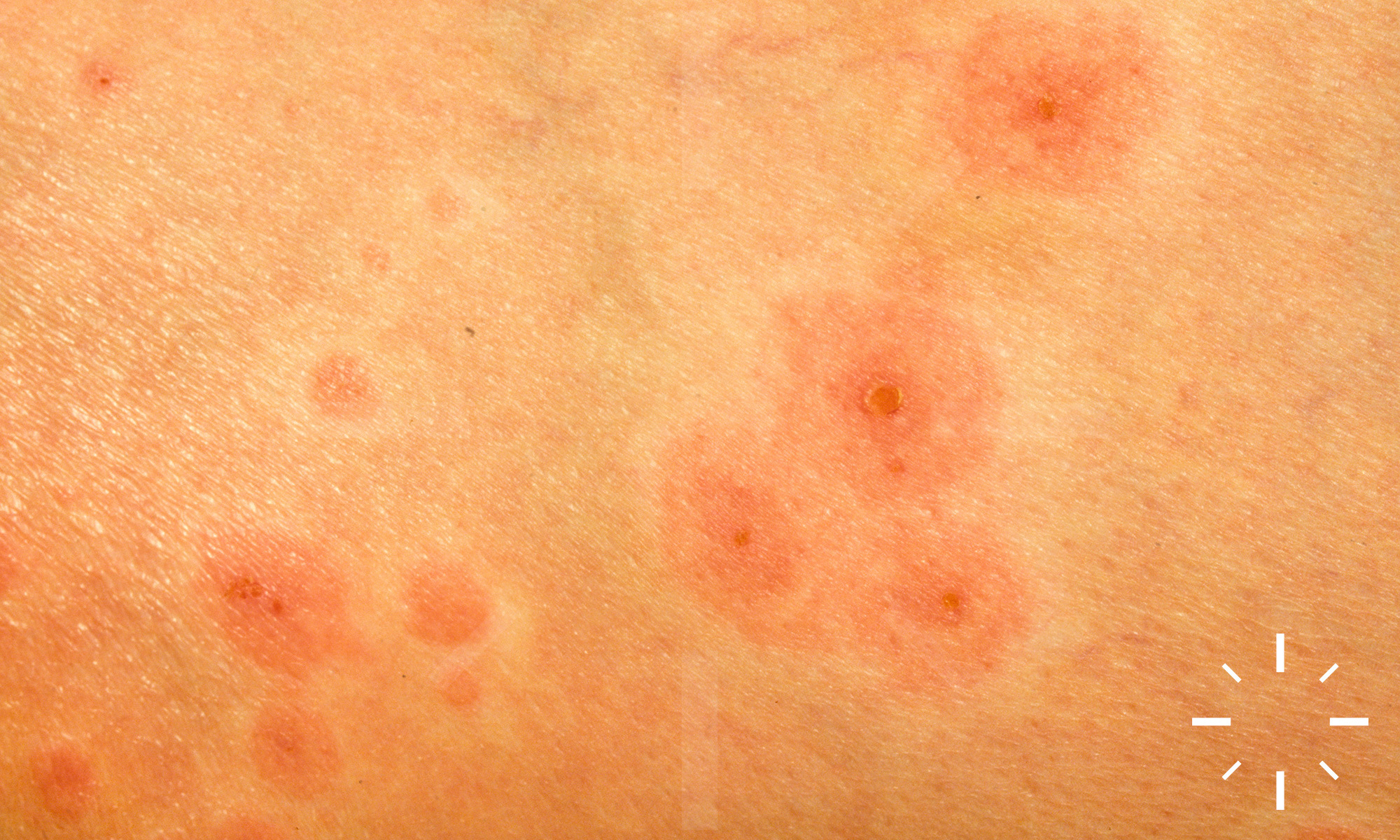
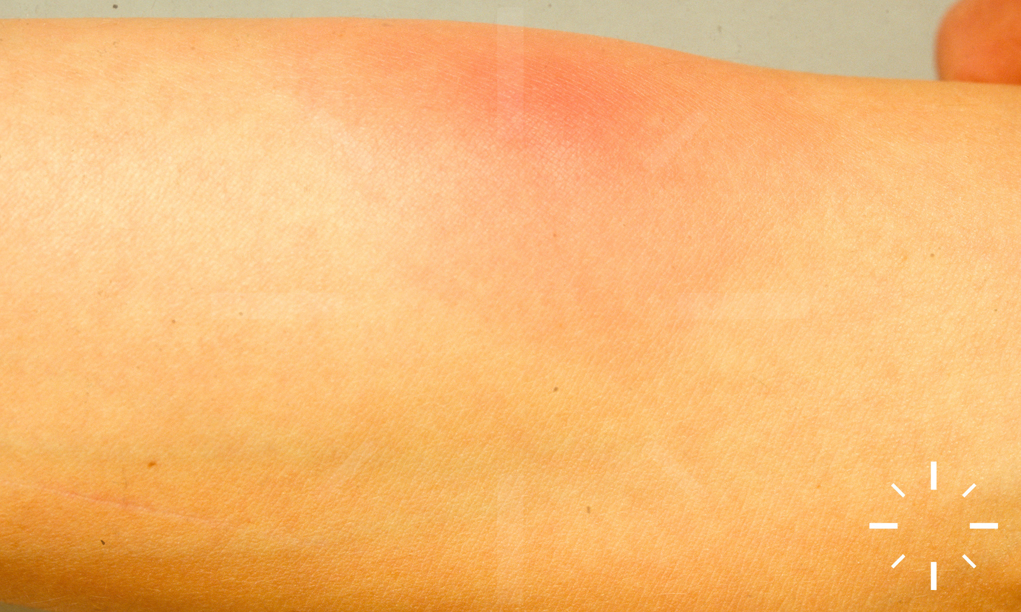
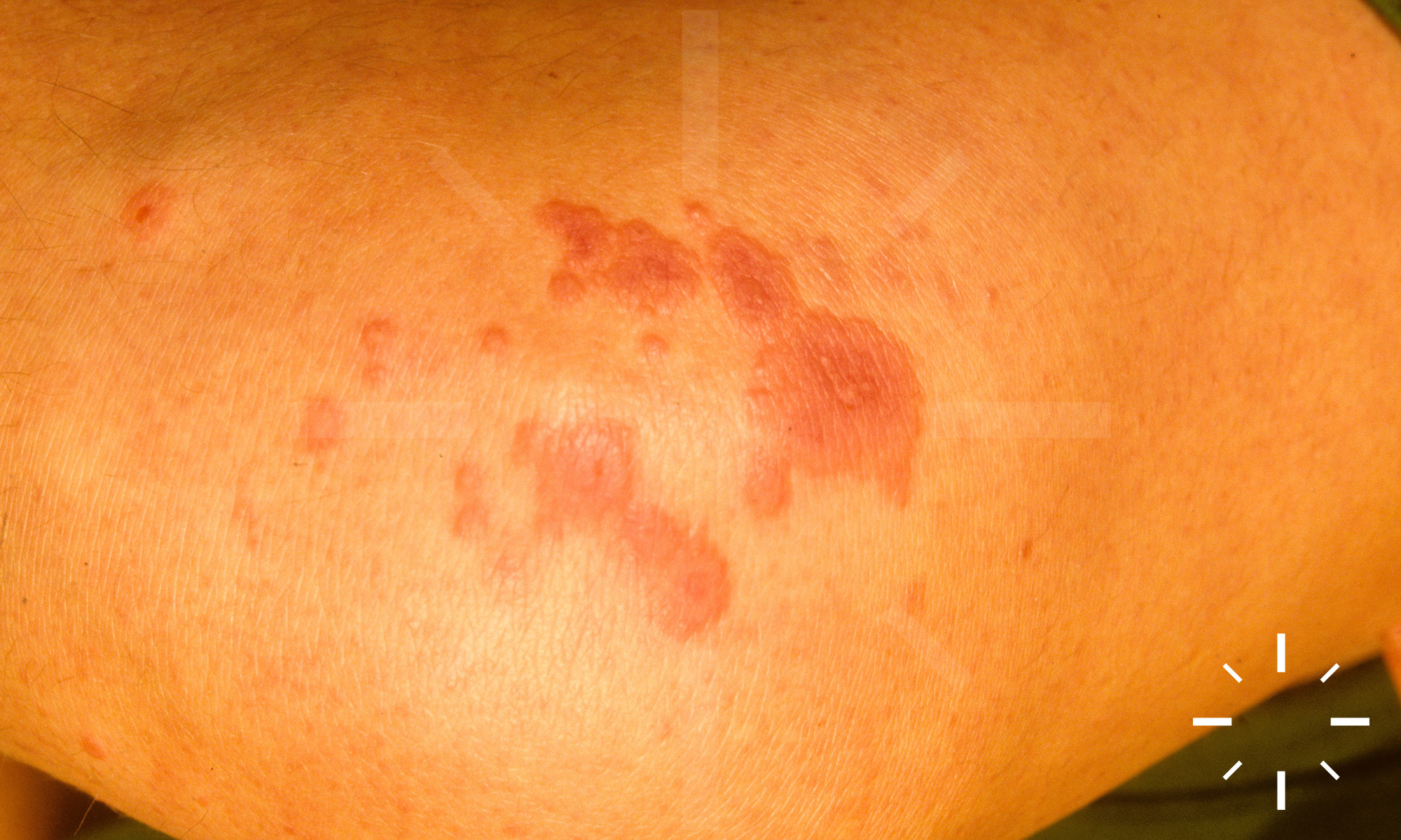
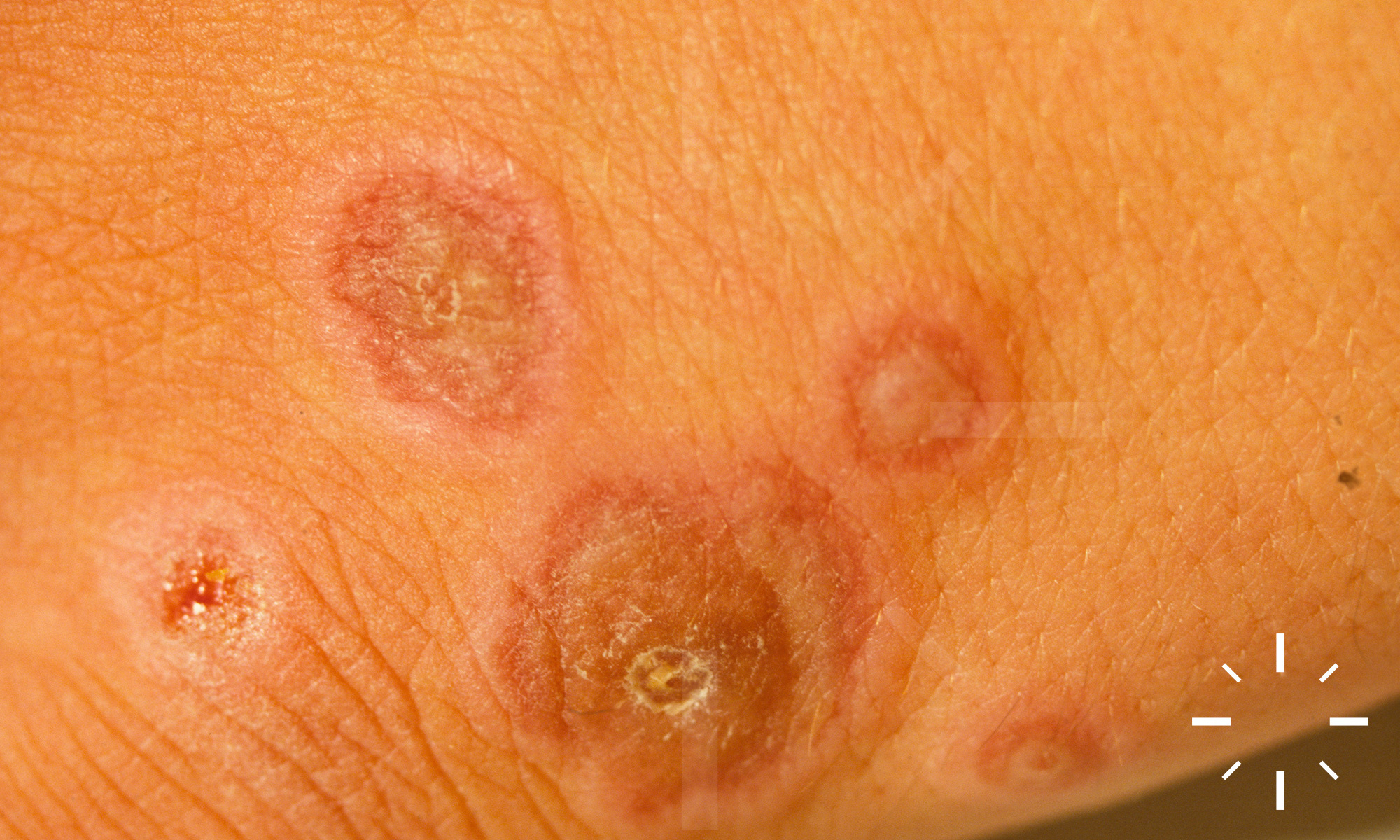
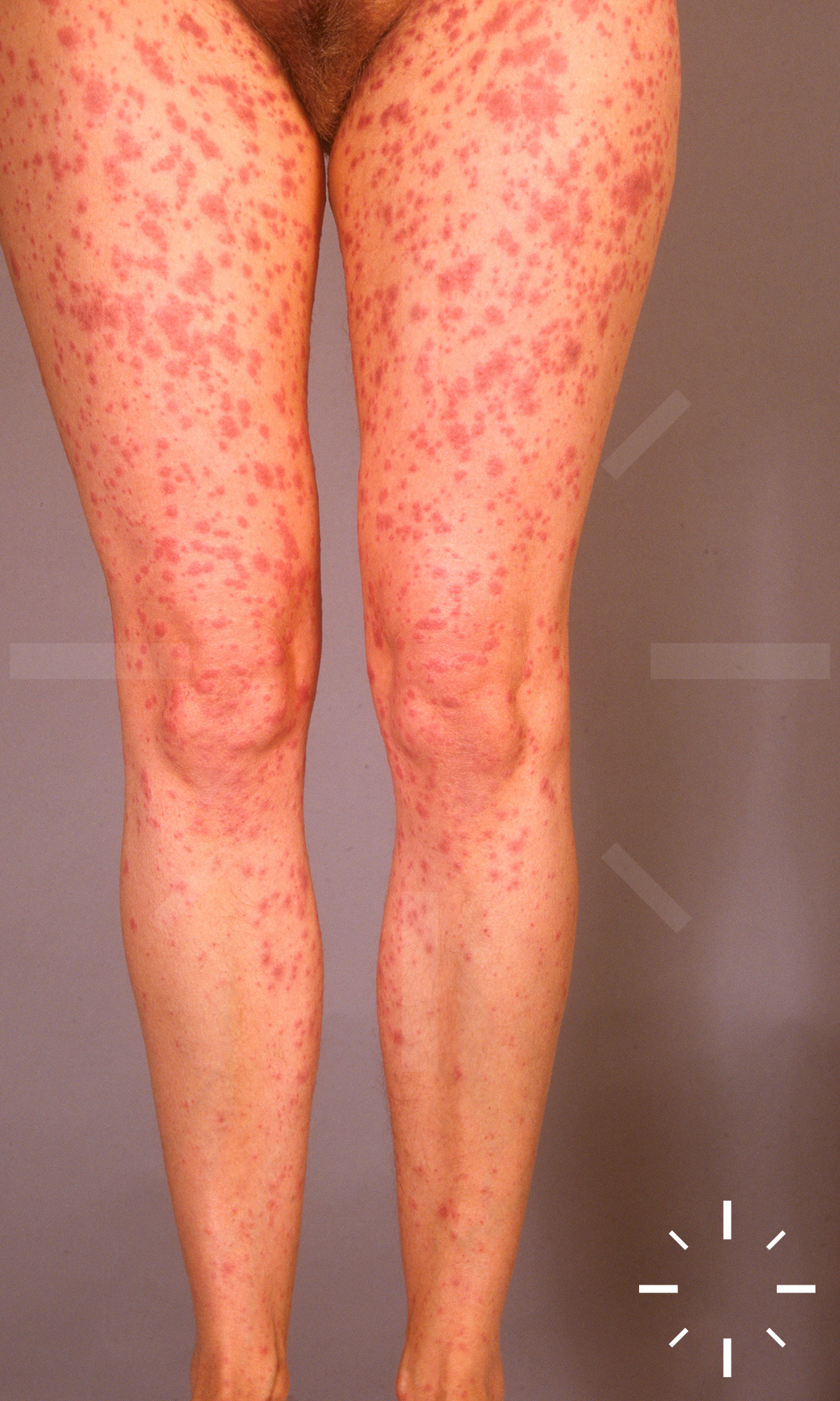
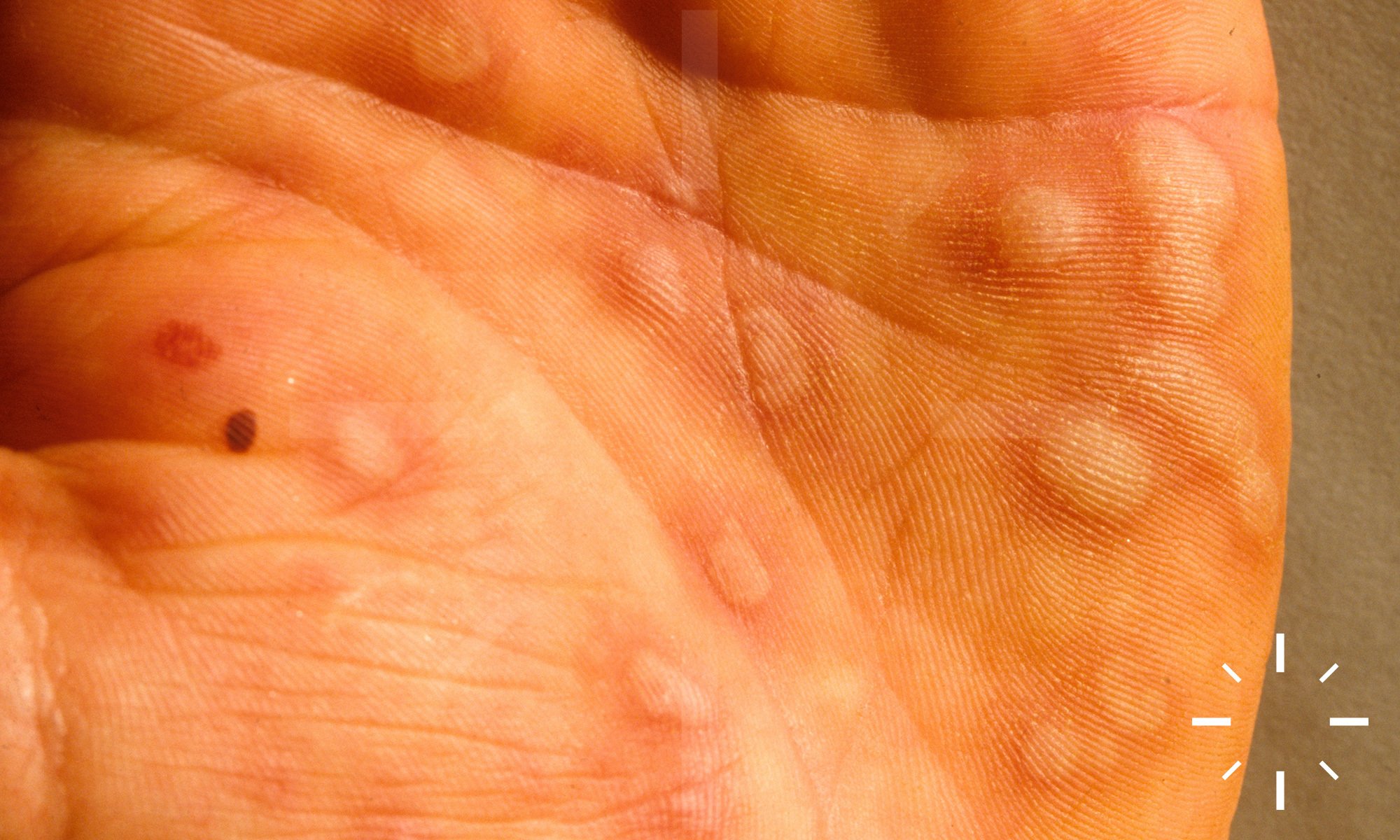
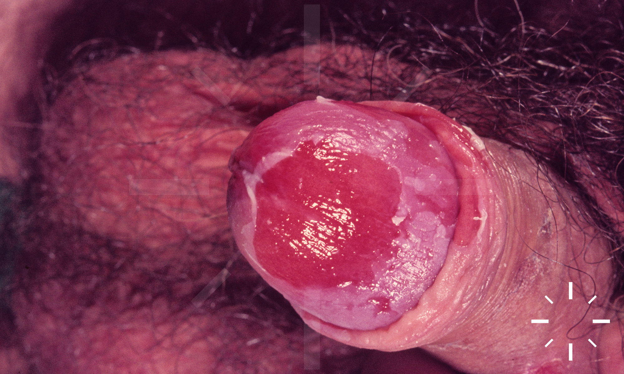
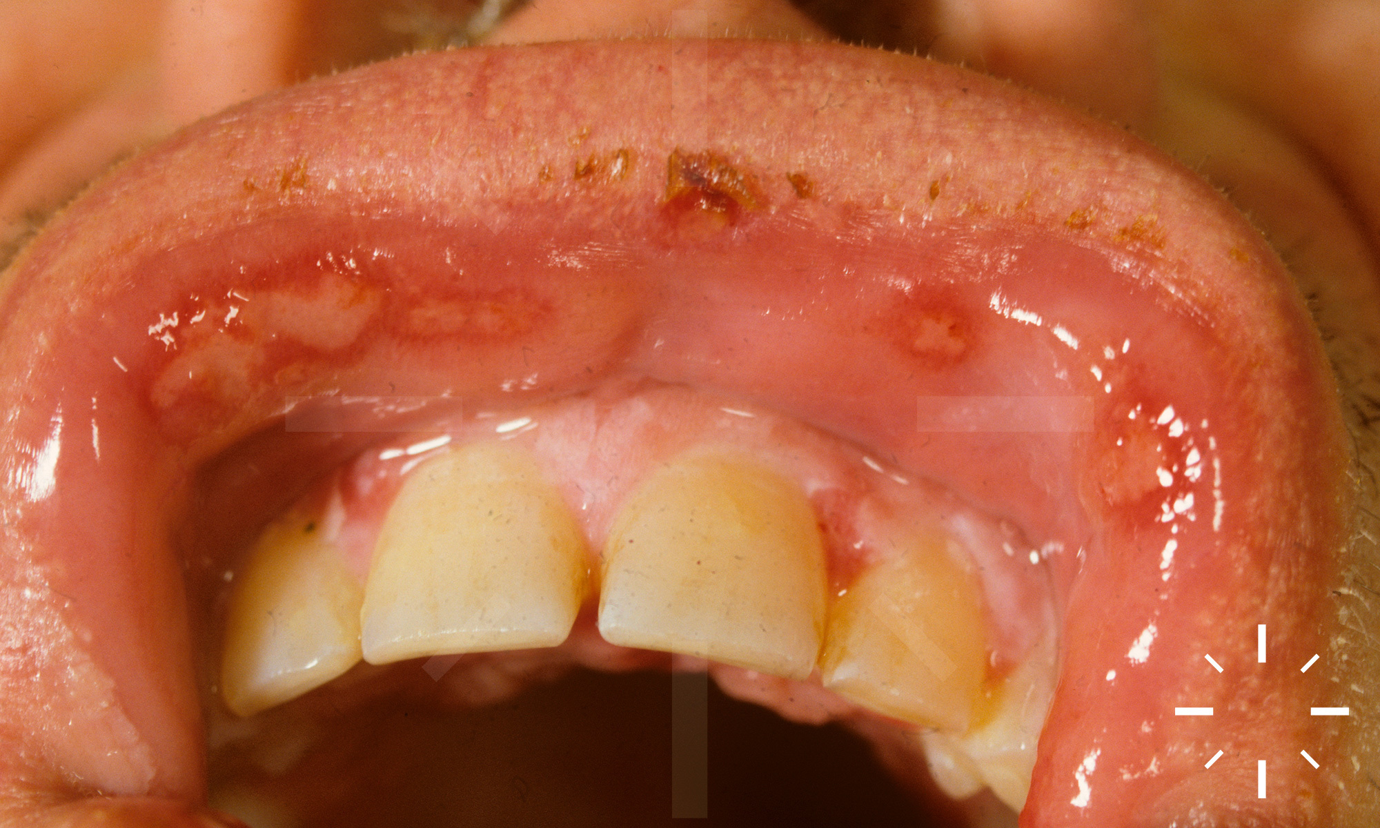
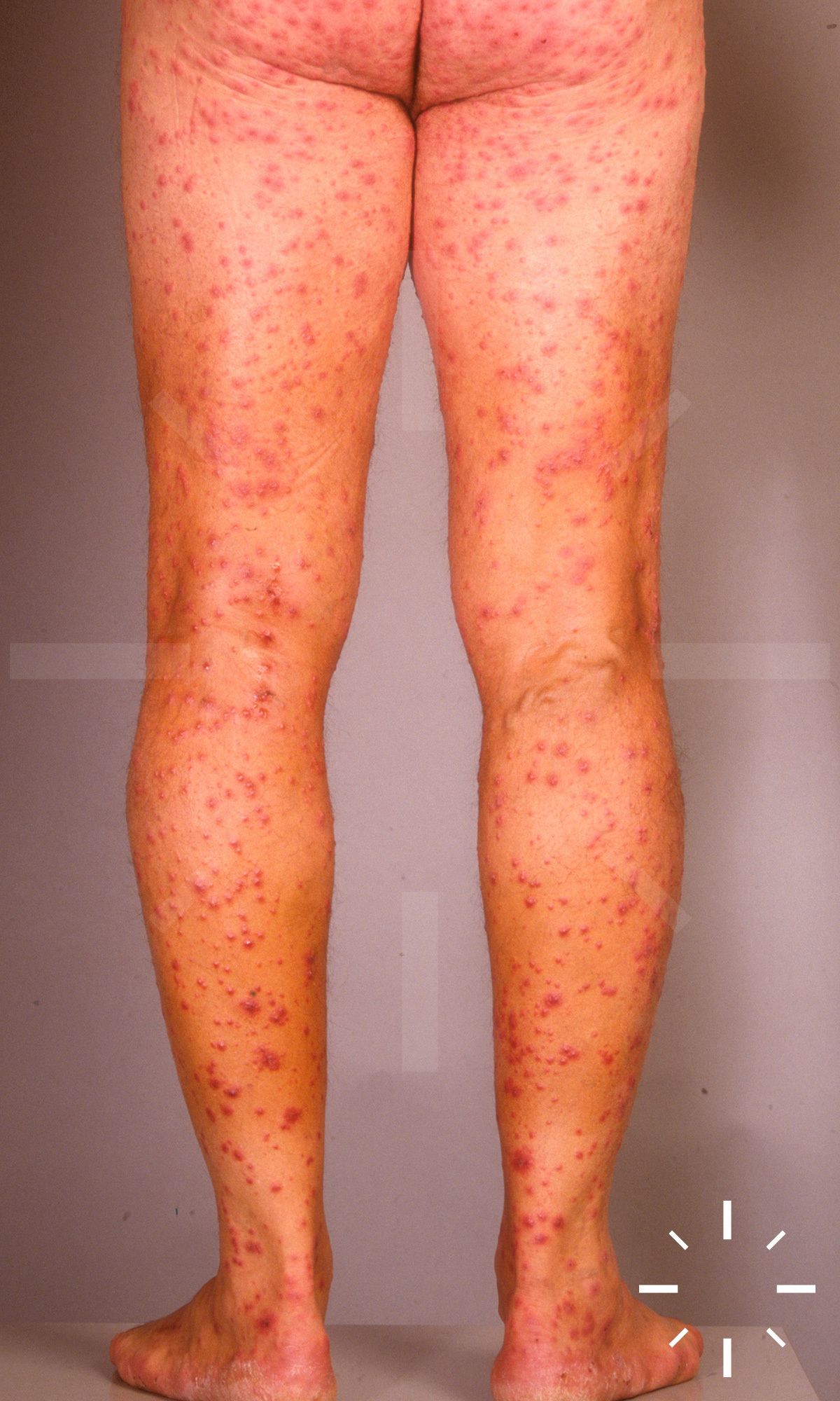
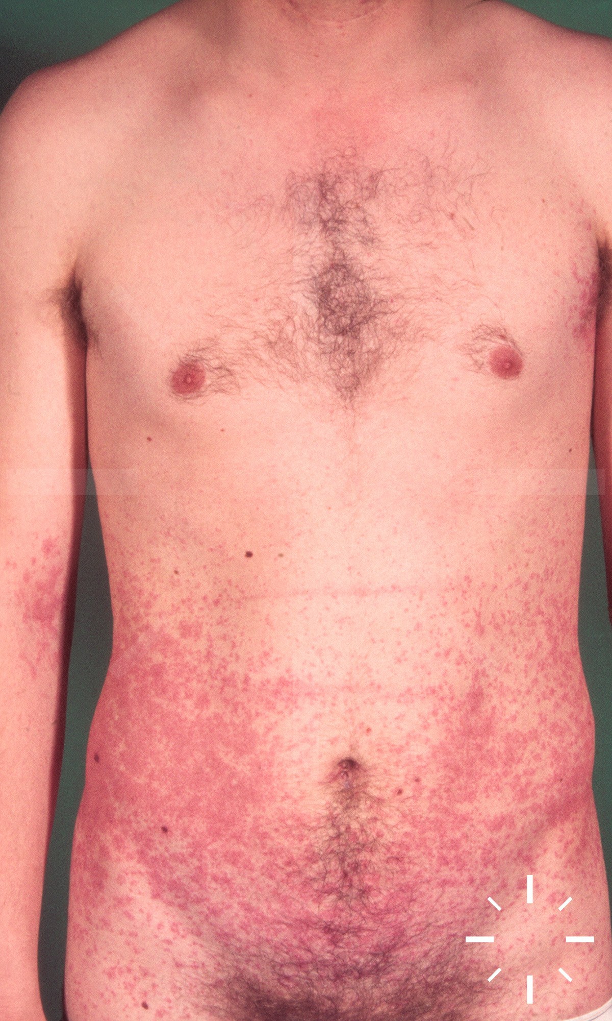
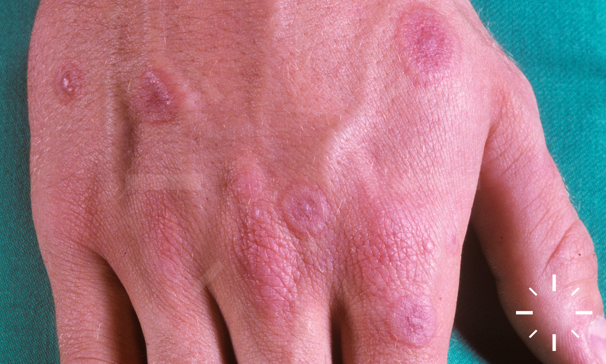
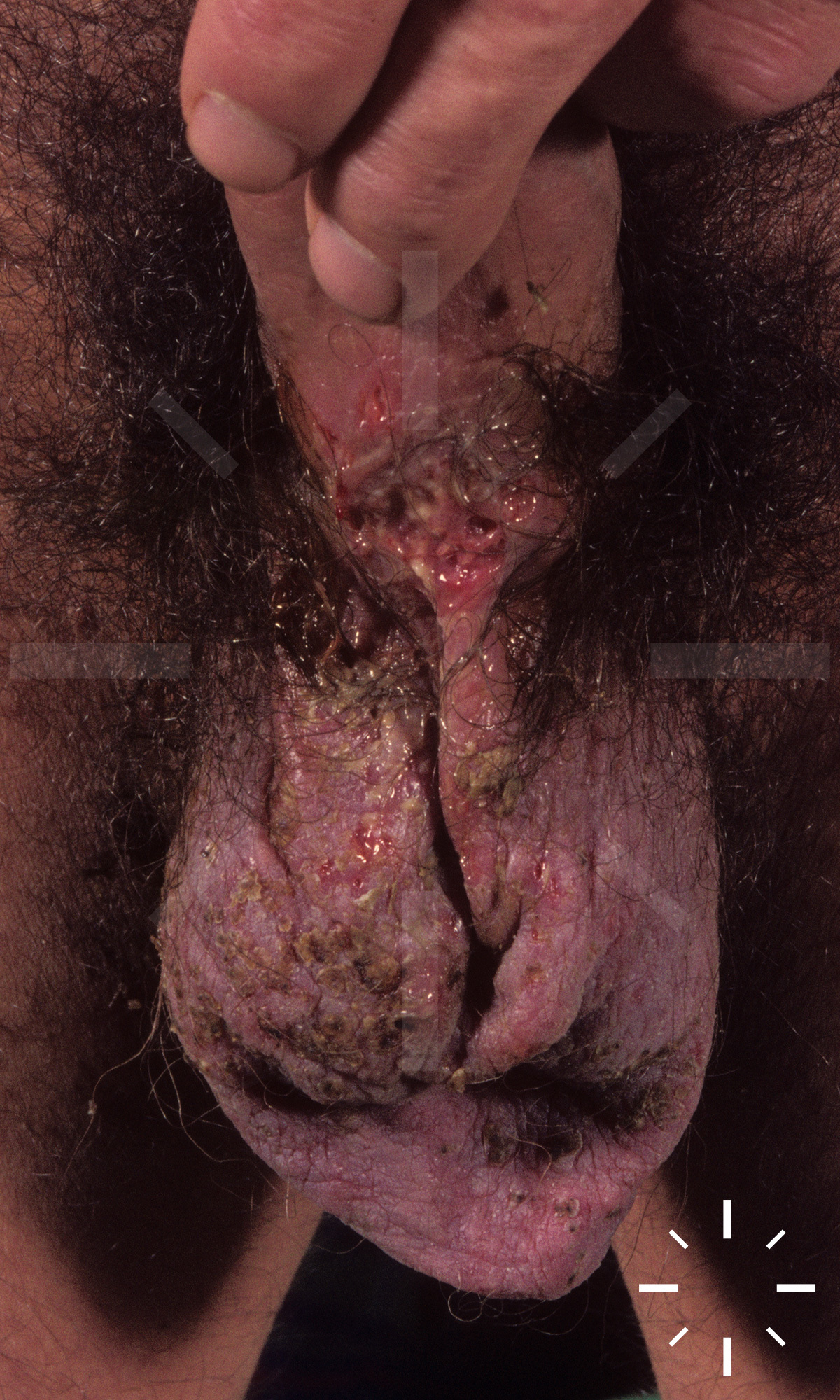
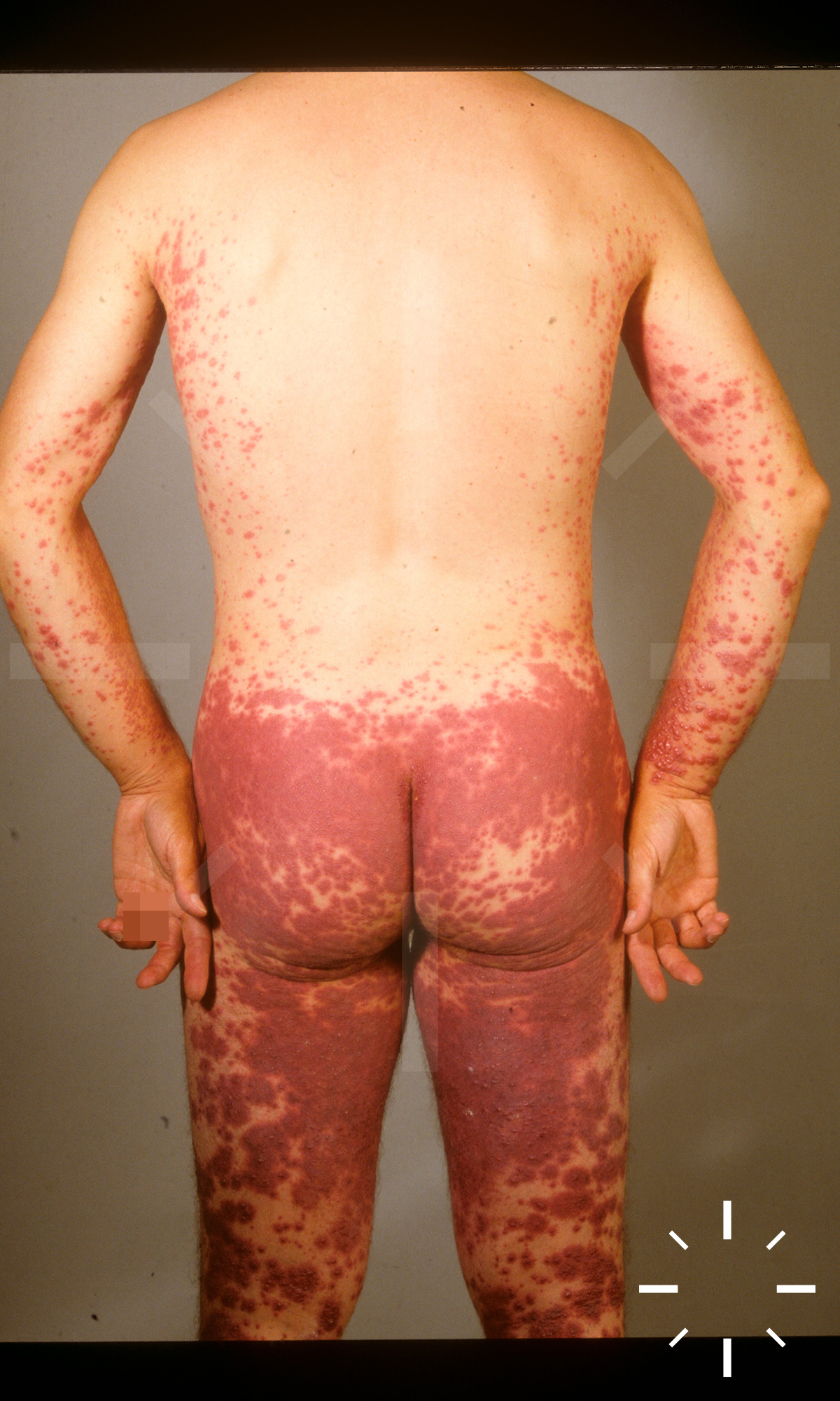
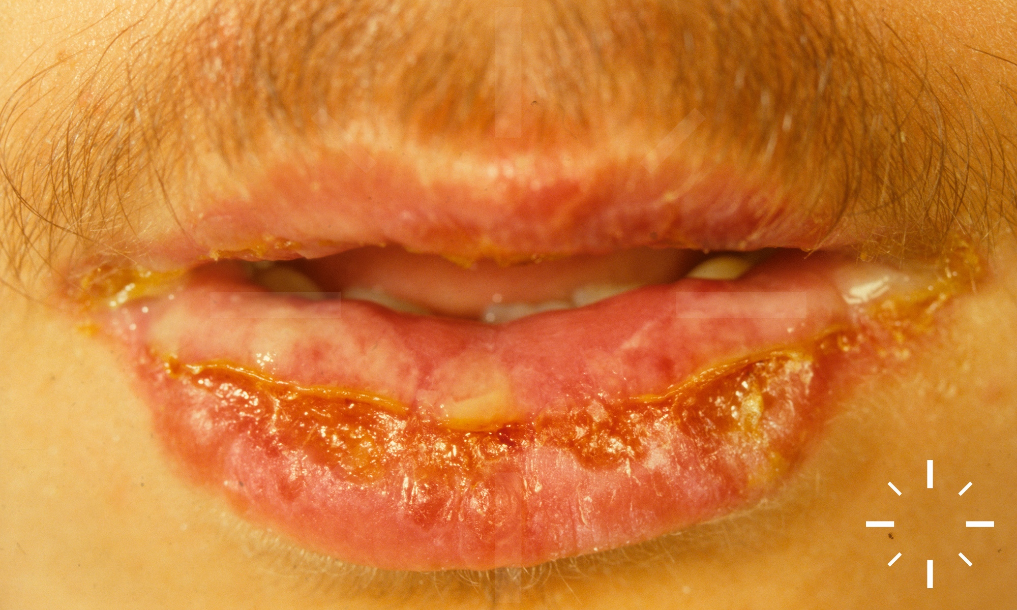
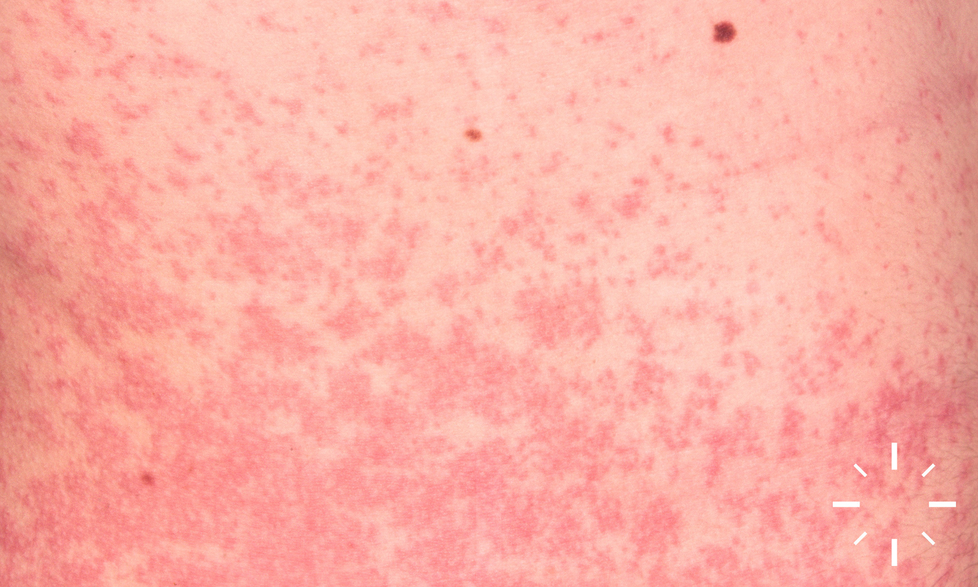
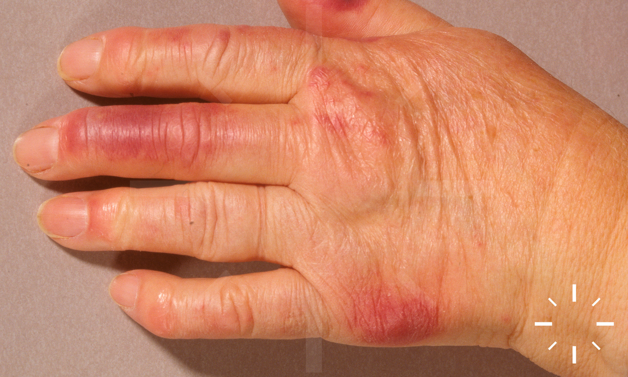
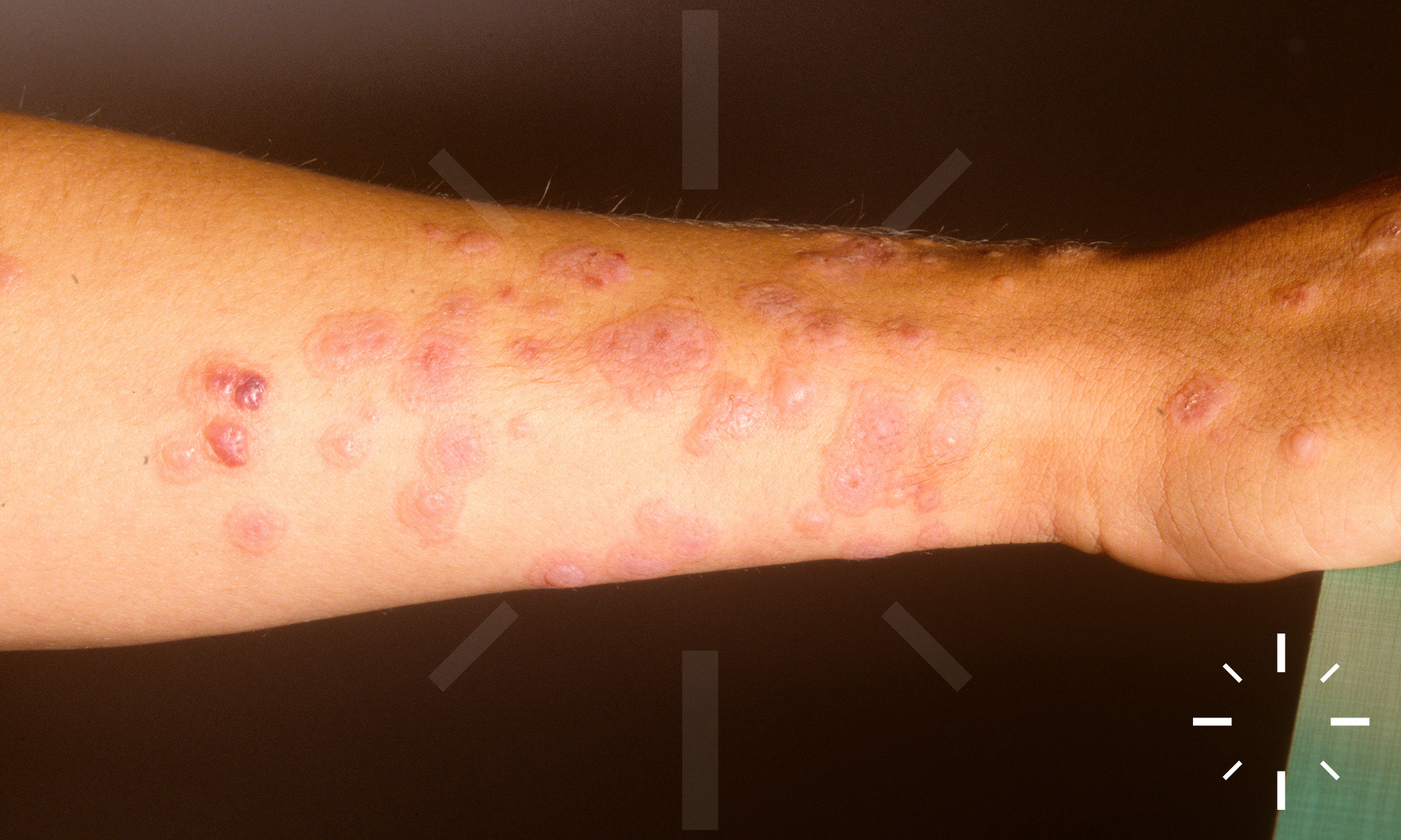
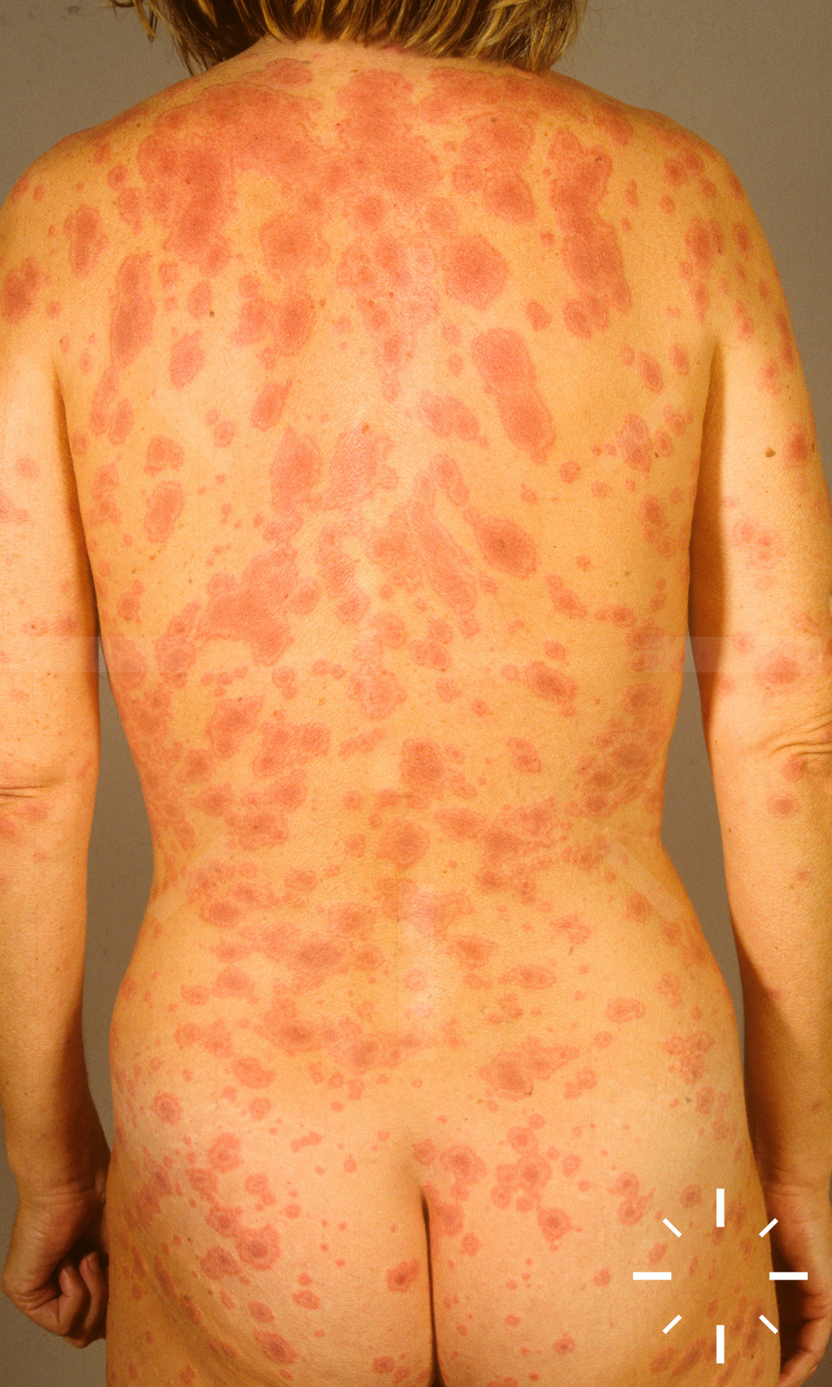
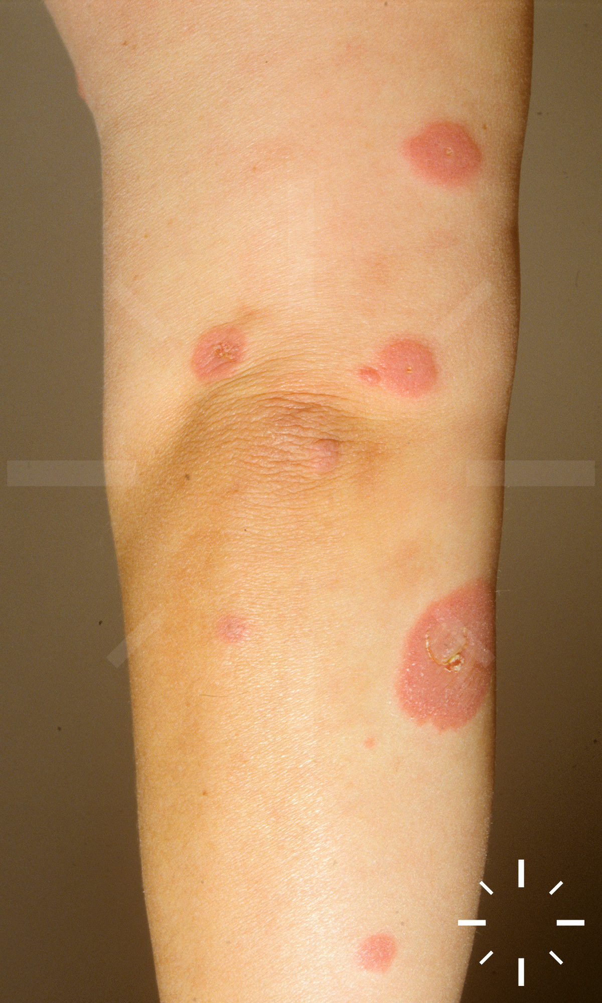
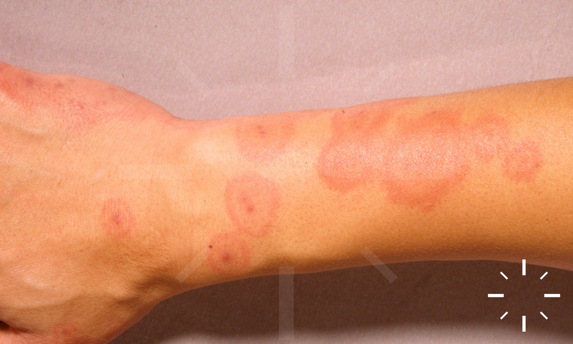
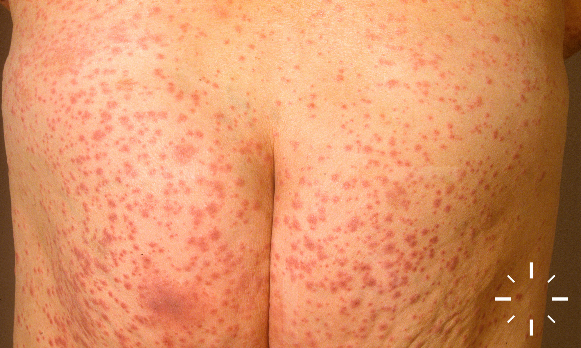
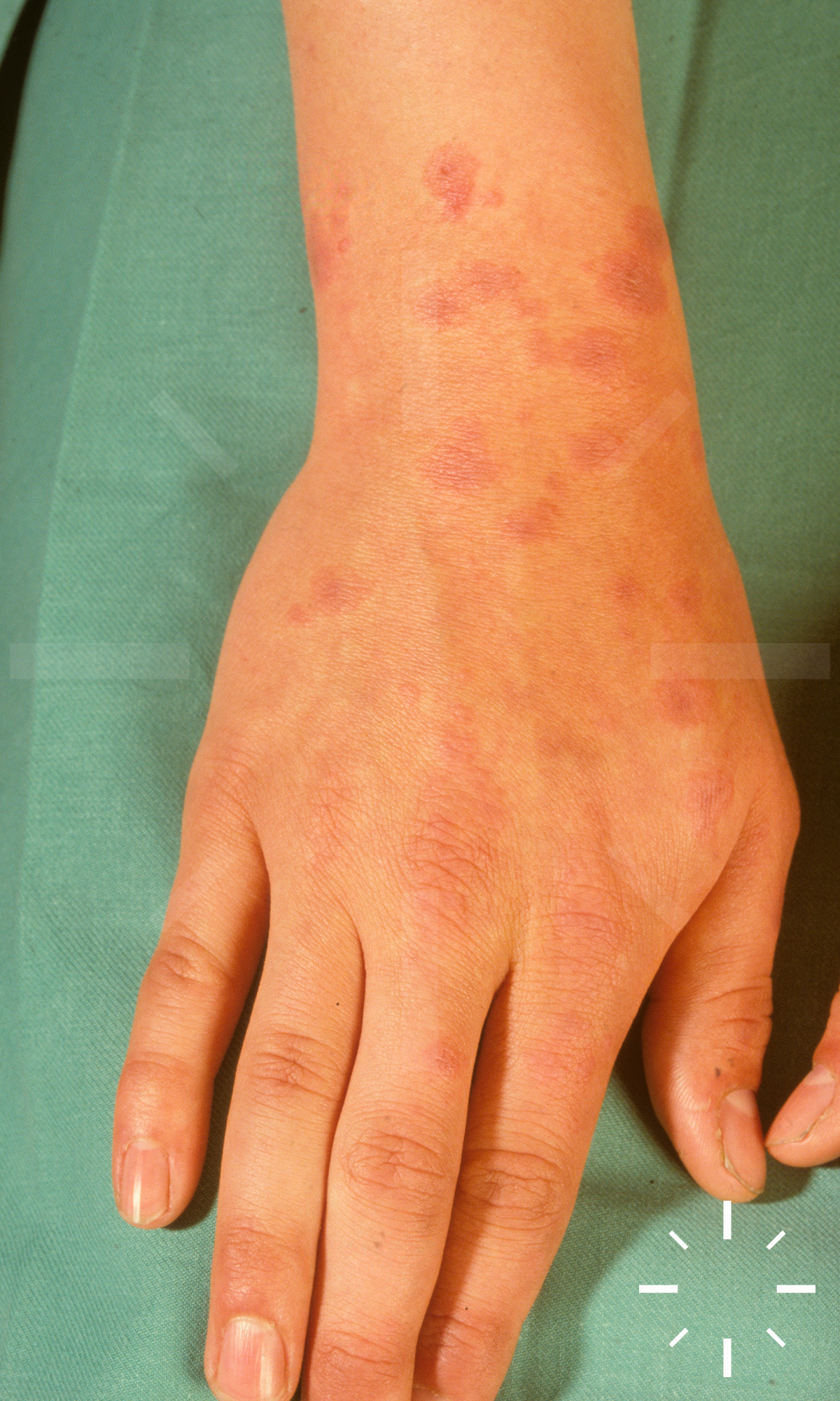
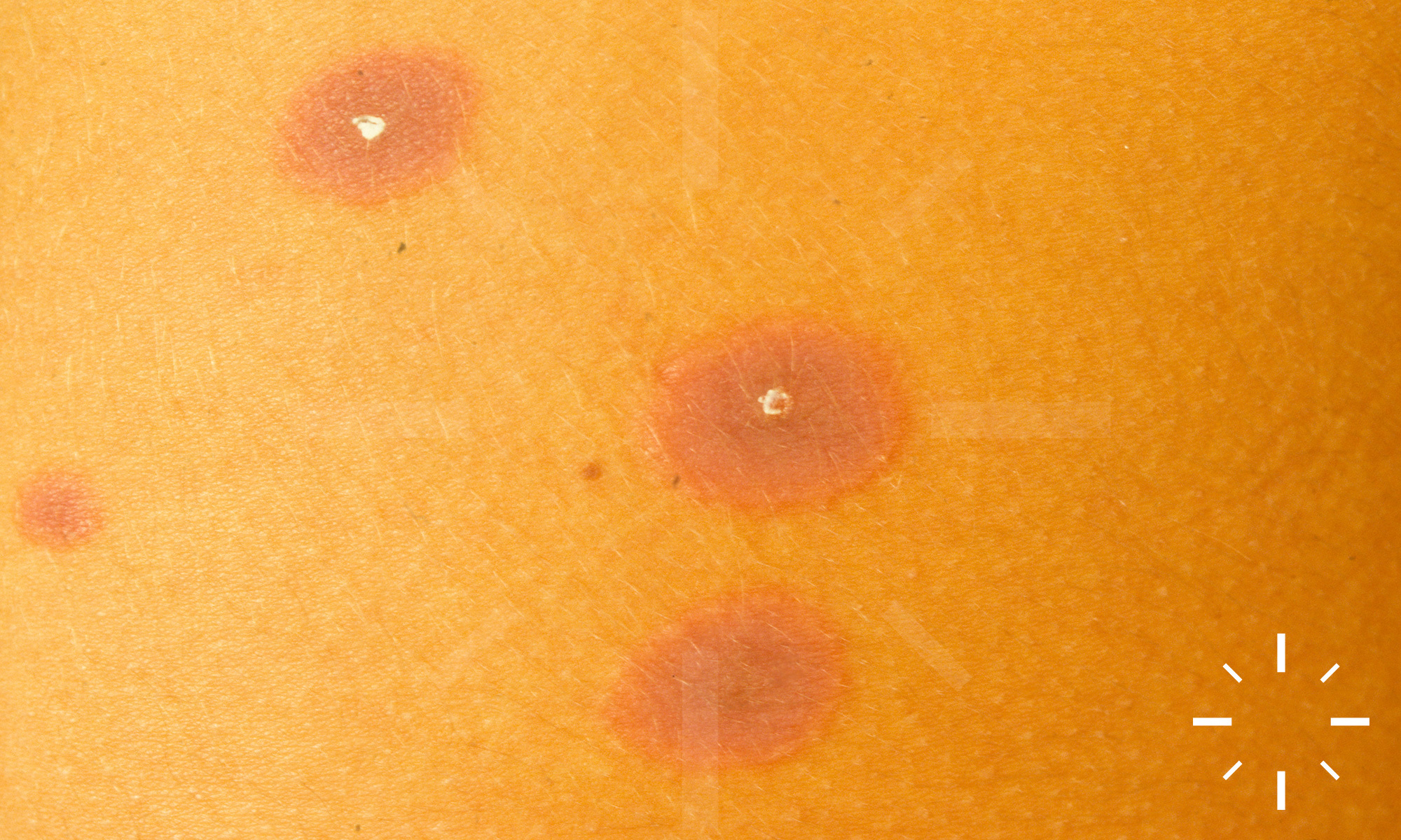
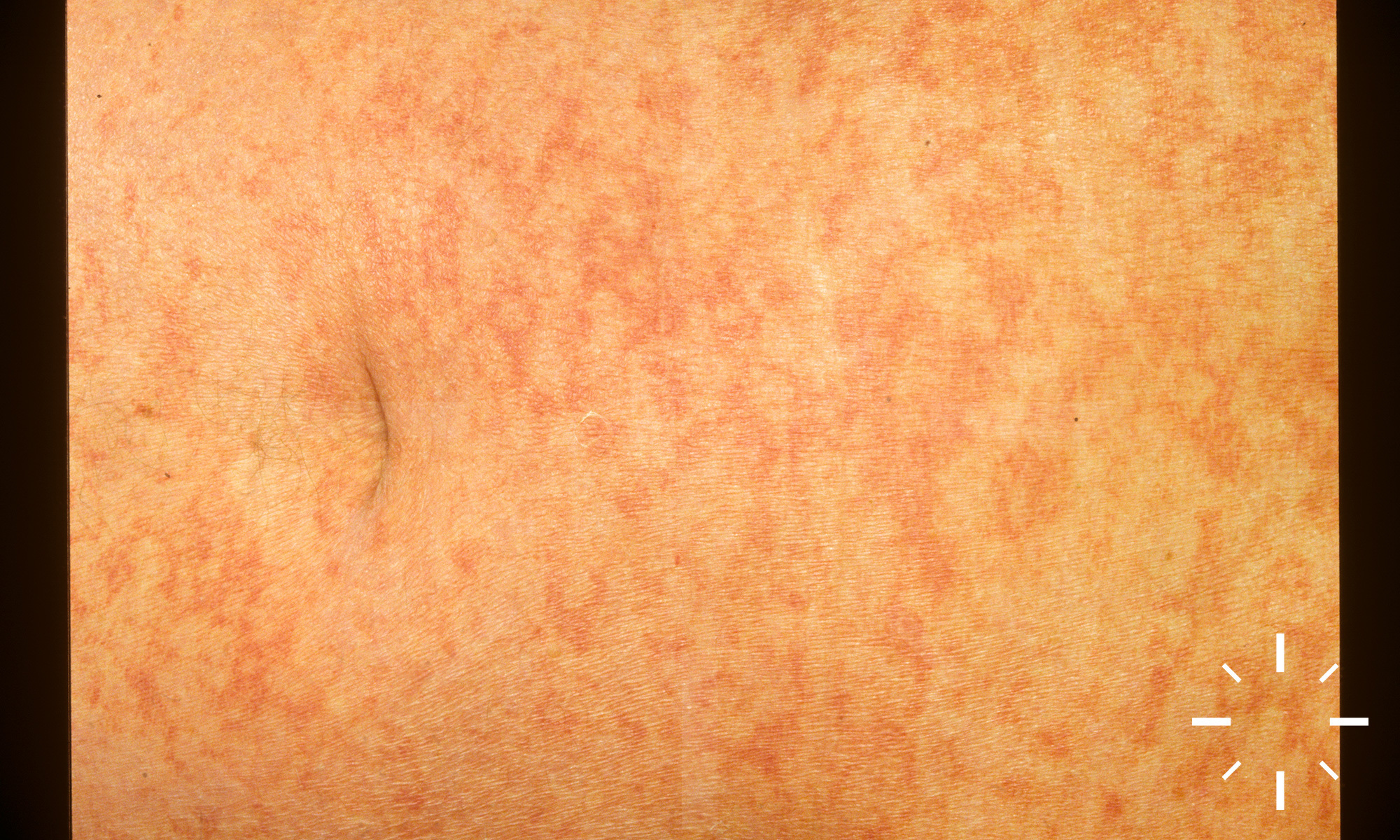
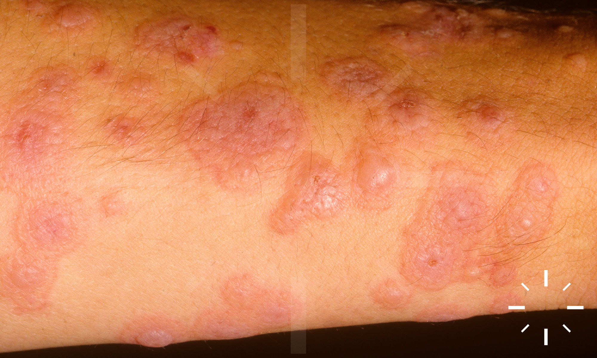
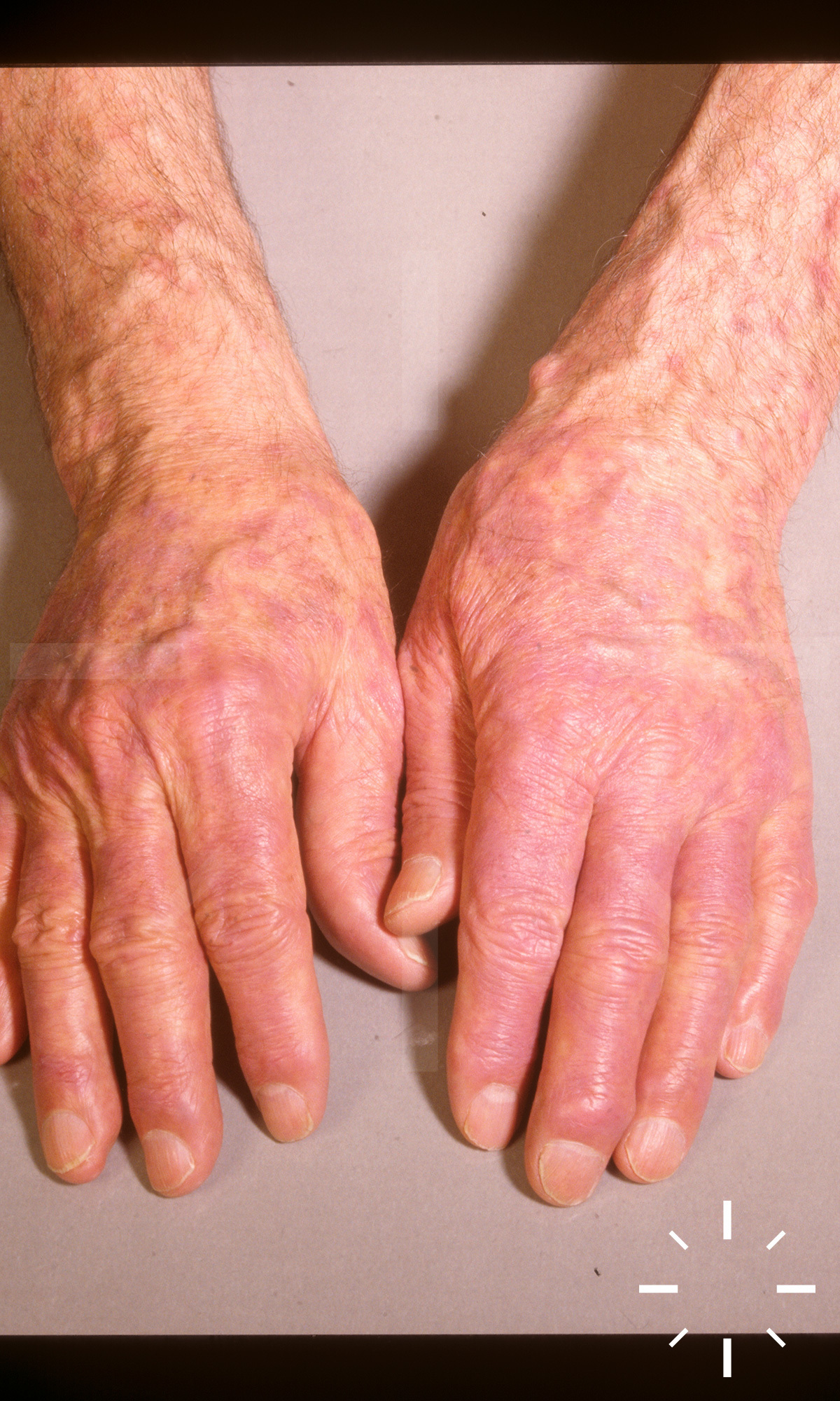
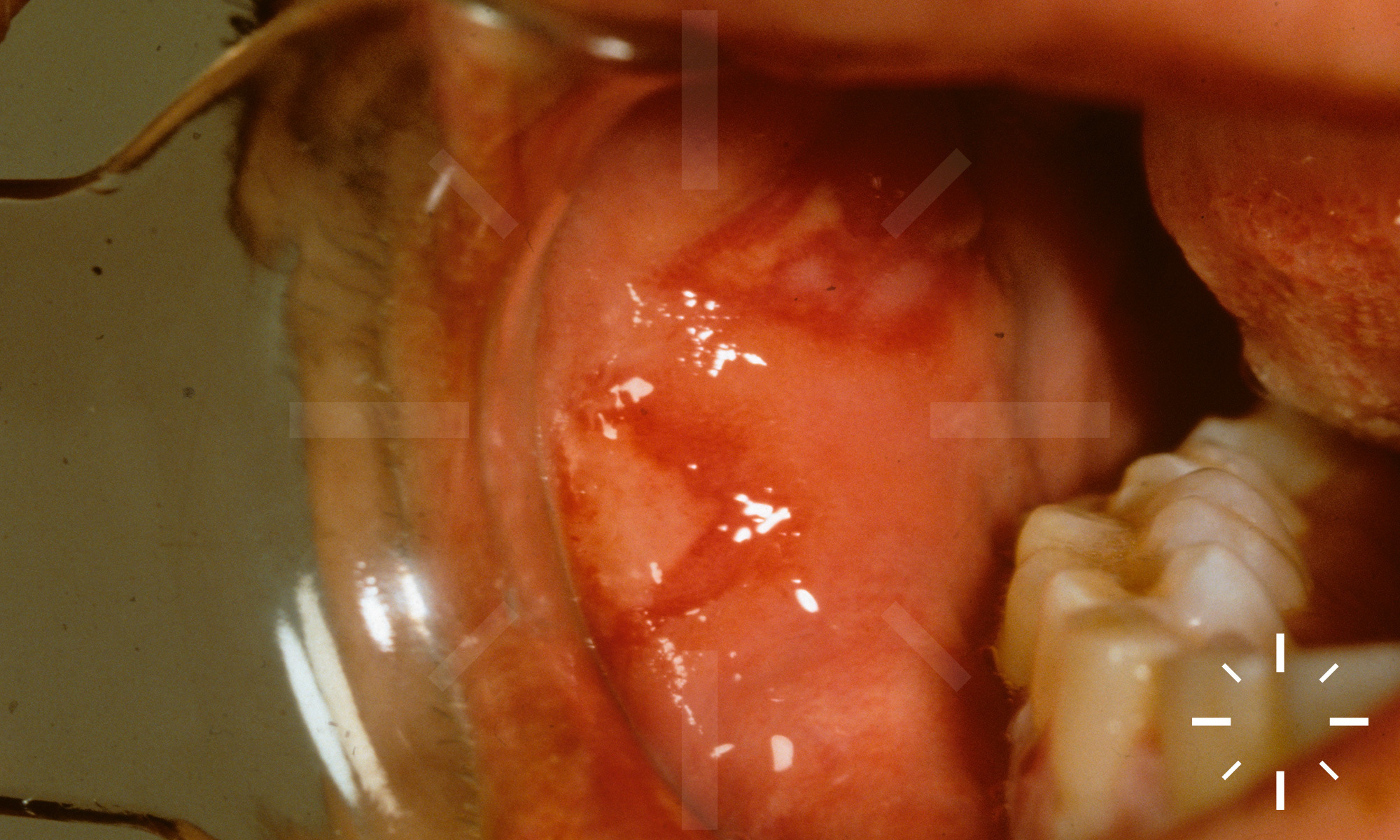
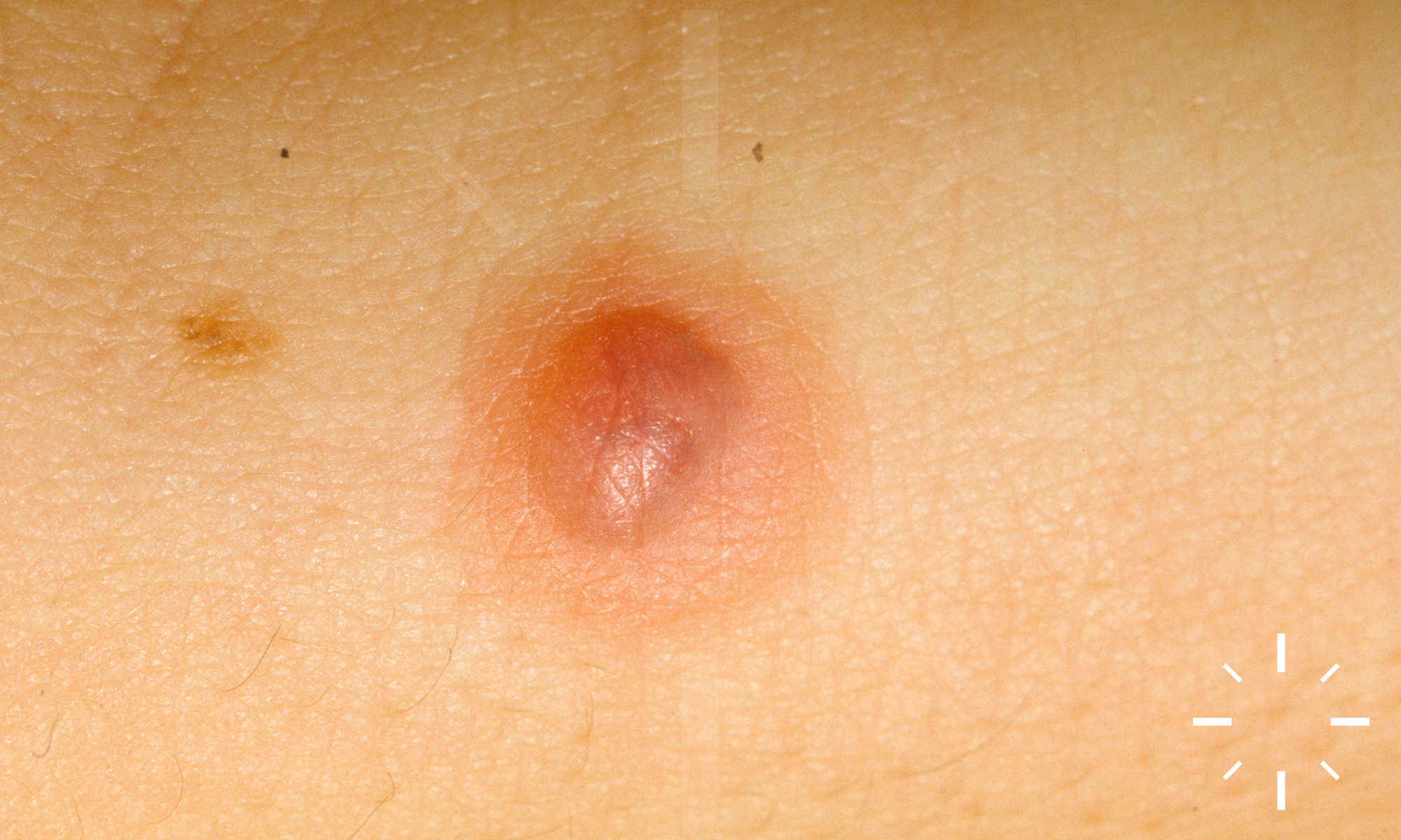
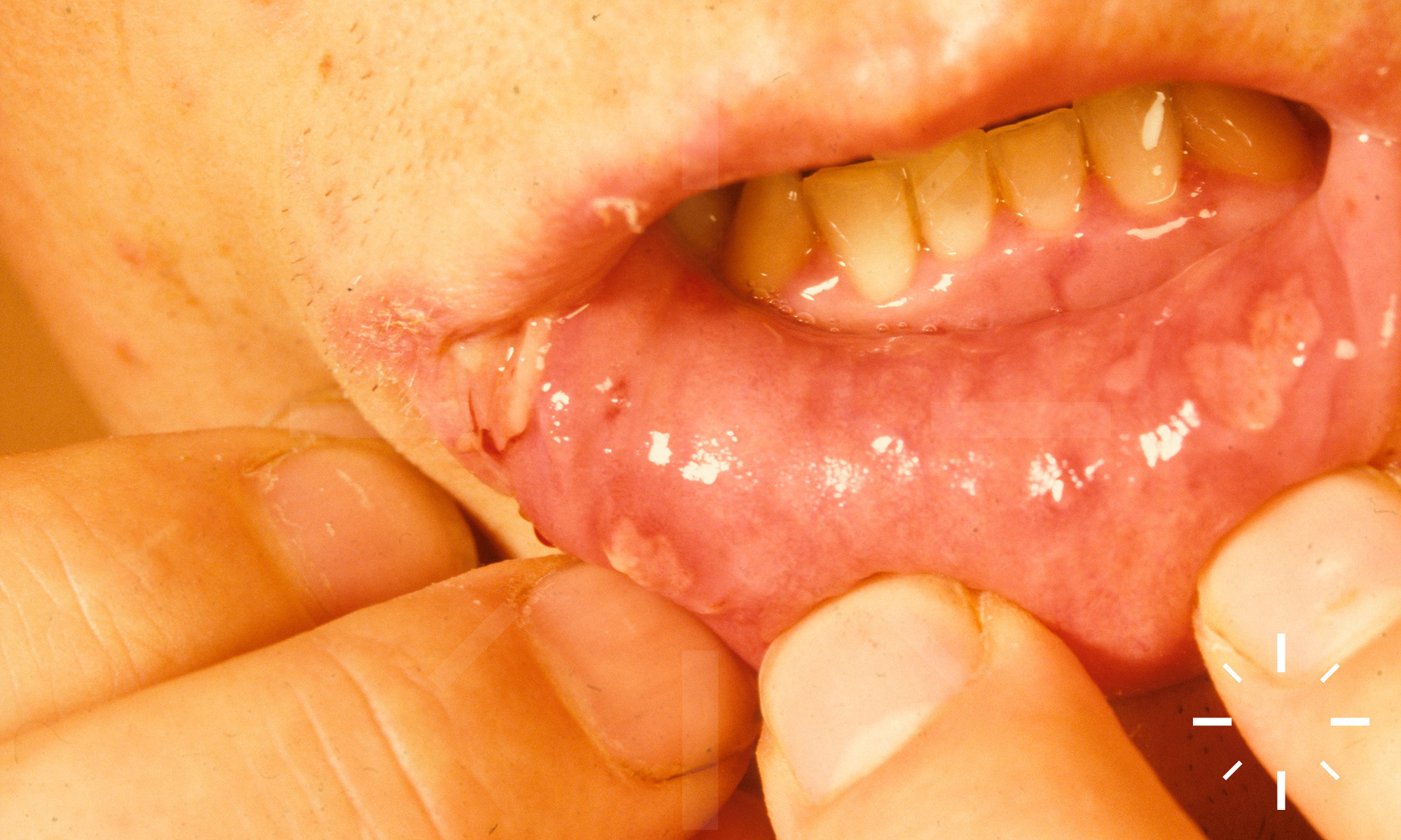
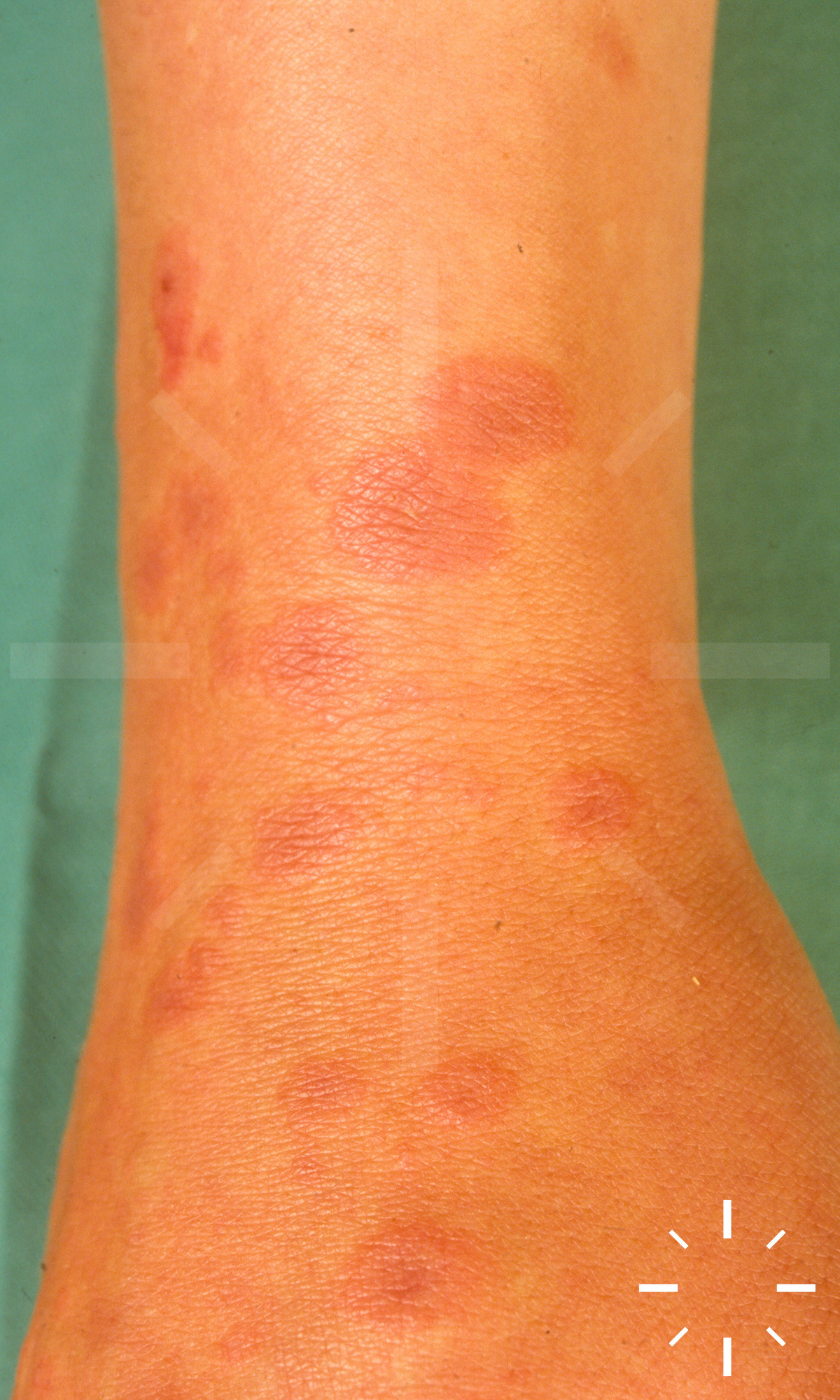
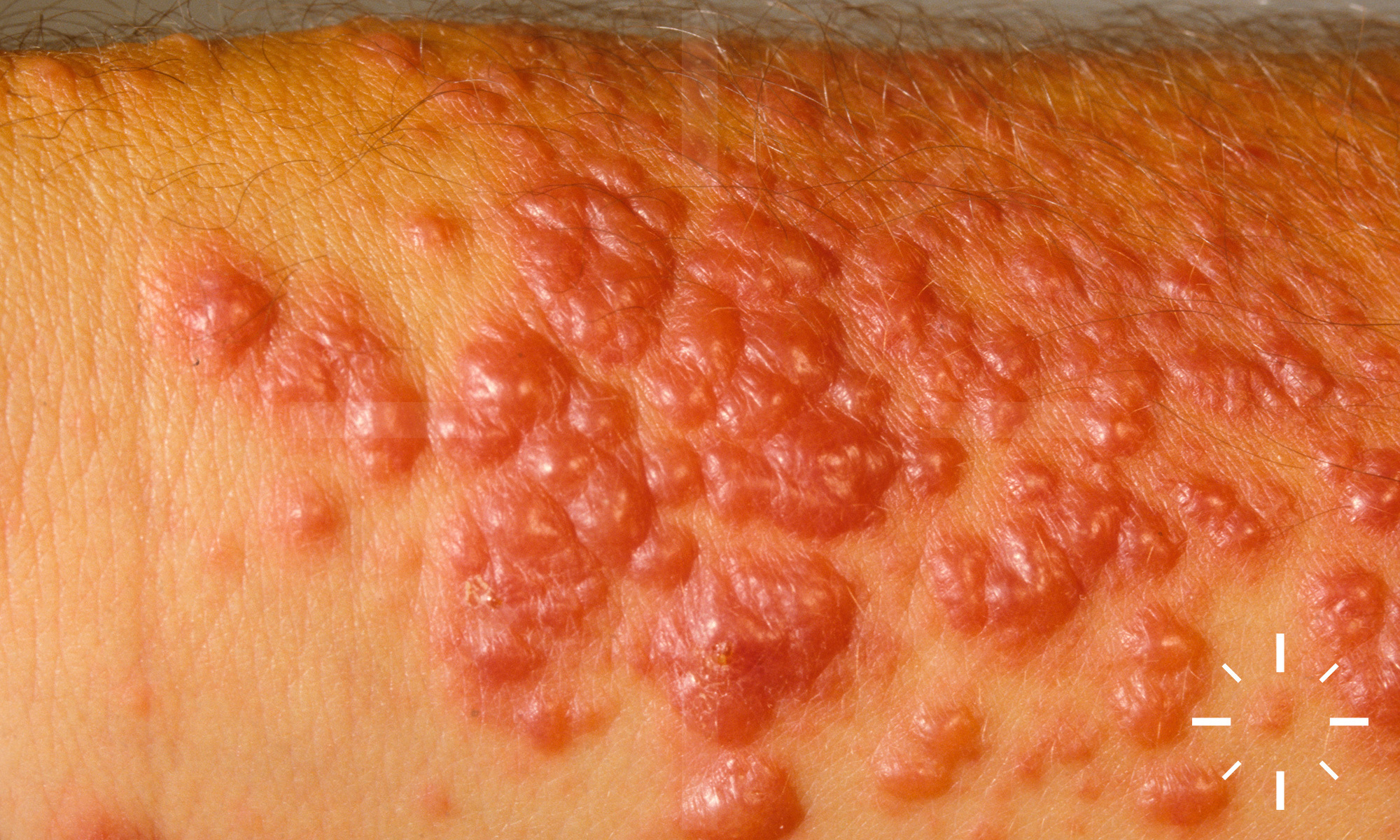
Erythema exsudativum multiforme

Last Updated: 2025-02-11

Author(s): Anzengruber F., Navarini A.

ICD11: EB12.Z

Ferdinand v. Hebra 1860

Erythema multiforme, EM, EEM, erythema multiforme of Hebra.

Acute, usually self-limiting, polyetiological disease characterised by target-like lesions.

  • Incidence: unknown, but estimated to be well below 1% per annum
  • Peak incidence: 20- 40 years of age
  • M > F

  • Minor form: no mucosal involvement. No systemic symptoms. No progression to toxic epidermal necrolysis (TEN).
  • Major form: with mucosal involvement. Systemic symptoms, usually before the skin changes: Fever and asthenia, occasionally arthralgias and atypical pneumonia. No progression to TEN. 

  • Possible triggers (combinations also possible)
  • Infections
    • approx. 90% of all triggers
    • Viral
      • Herpes simplex virus (HSV) type I (most common trigger)
      • HSV type II
      • Orf virus
      • Parapox virus
      • Varicella zoster virus
      • Epstein-Barr virus
      • Parvovirus B19
      • Adenovirus
      • Cytomegalovirus
      • Coxsackie virus
      • Hepatitis B or C virus
      • HI virus
    • Bacteria
      • Mycoplasma pneumoniae (frequent trigger, especially in children)
      • Chlamydophila psittaci
      • Salmonella
    • Mycobacteria
      • Mycobacterium tuberculosis
    • Mycoses
      • Histoplasma capsulatum
      • Dermatophytes
  • Medications (<10%)
    • NSAIDs
    • Sulfonamides
    • Antiepileptics
    • Antibiotics
  • Neoplasias
  • Autoimmune diseases
  • Vaccinations
  • UV-radiation (sunburn)
  • Trauma
  • Sarcoidosis
  • Menstruation
  • Possible genetic predisposition: HLA-DQw3, DRw53, AW33, HLA-DQB1*0301, B15(62), B35, and DQ3.
  • HSV infection causes cellular immune reactions against viral antigens localised in the skin.


  • EEM in general:

    • Typical target lesions: Two rings of contrasting erythema, smaller than 3cm, and central skin with signs of epidermal damage such as bulla or crusting.

    • There are usually erythematous, often pruritic or burning papules at localised sites, which transform exanthematously into cocardial and disc-shaped plaques with characteristic central vesicles. A linear arrangement may be detectable (Köbner phenomenon), but this is only active BEFORE the EEM.

    • The first skin changes usually appear after an incubation period of 2-3 days. In some cases, however, there can be up to 17 days between contact with the trigger and the first appearance of efflorescences.

  • Prodromal symptoms can occur with EEM major:

    • ↓ AZ, fever, myalgias

    • Cough with M. pneumoniae infection

    • EEM major: Enanthema, painful erosions or vesicles appear on the mucous membranes. Usually only the oral cavity is affected, but in a few cases it can lead to involvement of the pharynx and upper respiratory organs.  In most cases, only the oral mucosa is affected. Involvement of the genital mucosa occurs in approx. 25% and ocular involvement in approx. 17%.

  • The most important differential diagnosis is Steven Johnson syndrome: it produces dark red macules and atypical targets, as well as bullous lesions, and can develop into TEN with epidermal detachment of >30% of the body surface area.

  • Most common false differential diagnosis is annular urticaria gigantea, also known as urticaria multiforme. Attention, EM lesions remain in place for 7 days, but urticaria only for 24 hours. Subcutaneous adrenaline does not affect EEM lesions, but urticaria disappears in 30 minutes. 

  • Anamnesis (infections, herpes simplex, cough, medication intake)
  • Clinic
  • Laboratory:
    • Nonspecific
    • Optional: ↑BSG, leucocytosis, ↑transaminases, HSV PCR
  • Urine
    • Mycoplasma pneumoniae
  • Biopsy
    • Dermatopathology
    • Direct immunofluorescence

EEM minor: Mostly symmetrical on the extensor sides of the extremities, palmoplantar, elbows, face.

EEM major: Face, extremities and mucous membranes can be affected. The spread is centripetal.

Focal apoptosis of keratinocytes with interface dermatitis. Vacuolar degeneration of the stratum basale. Lymphocytic, perivascular infiltrates. In the advanced stage, confluent keratinocyte necrosis occurs.

Normally without sequelae. Eye damage with EEM major is rare.

In HSV-triggered EEM, a continuous HSV prophylaxis with valaciclovir can be indicated.

  • Scarless healing, but in some cases with hyperpigmentation, within 2 weeks.
  • Very few patients suffer from recurrences, some up to 6 times a year. The average duration of recurrences is 6-10 years.

  1. Aslanzadeh J, Helm KF, Espy MJ, Muller SA , Smith TF. Detection of HSV-specific DNA in biopsy tissue of patients with erythema multiforme by polymerase chain reaction. Br J Dermatol 1992;126:19-23.
  2. Assier H. Erythema Multiforme With Mucous Membrane Involvement and Stevens-Johnson Syndrome Are Clinically Different Disorders-Reply. Arch Dermatol 1996;132:711.
  3. Atzori L, Pau M , Aste M. Erythema multiforme ID reaction in atypical dermatophytosis: a case report. Journal of the European Academy of Dermatology and Venereology 2003;17:699-701.
  4. Auquier-Dunant A, Mockenhaupt M, Naldi L, Correia O, Schröder W , Roujeau J-C. Correlations Between Clinical Patterns and Causes of Erythema Multiforme Majus, Stevens-Johnson Syndrome, and Toxic Epidermal Necrolysis. Arch Dermatol 2002;138.
  5. Bastuji-Garin S. Clinical Classification of Cases of Toxic Epidermal Necrolysis, Stevens-Johnson Syndrome, and Erythema Multiforme. Arch Dermatol 1993;129:92.
  6. Batalla A, Alvarez-Arguelles ME, Gonzalez-Martinez MB , Curto JR. [Milker's nodule complicated with erythema multiforme]. Med Clin (Barc) 2013;141:e5.
  7. Brice SL, Leahy MA, Ong L, Krecji S, Stockert SS, Clark Huff J et al. Examination of non-involved skin, previously involved skin, and peripheral blood for herpes simplex virus DNA in patients with recurrent herpes-associated erythema multiforme. J Cutan Pathol 1994;21:408-12.
  8. Clark Huff J , Weston WL. Isomorphic phenomenon in erythema multiforme. Clin Exp Dermatol 1983;8:409-13.
  9. Côté B. Clinicopathologic Correlation in Erythema Multiforme and Stevens-Johnson Syndrome. Arch Dermatol 1995;131:1268.
  10. Darragh TM, Egbert BM, Berger TG , Benedict Yen TS. Identification of herpes simplex virus DNA in lesions of erythema multiforme by the polymerase chain reaction. Journal of the American Academy of Dermatology 1991;24:23-6.
  11. Dikiand WJ, Oranje AP, Stoiz E , Joost T. Erythema Multiforme in Childhood and Early Infancy. Pediatric Dermatology 1986;3:135-9.
  12. Farthing PM, Maragou P, Coates M, Tatnall F, Leigh IM , Williams DM. Characteristics of the oral lesions in patients with cutaneous recurrent erythema multiforme. J Oral Pathol Med 1995;24:9-13.
  13. Howland WW, Golttz LE, Weston WL , Huff JC. Erythema multiforme: Clinical, histopathologic, and immunologic study. Journal of the American Academy of Dermatology 1984;10:438-46.
  14. Huff JC. Erythema multiforme. Dermatol Clin 1985;3:141-52.
  15. Huff JC , Weston WL. Recurrent erythema multiforme. Medicine 1989;68:133-40.
  16. Johnston GA, Ghura HS, Carter E , Graham-Brown RAC. Neonatal erythema multiforme major. Clin Exp Dermatol 2002;27:661-4.
  17. Khalil I, Lepage V, Douay C, Morin L, Al-Daccak R, Wallach D et al. HLA DQB1*0301 Allele is Involved in the Susceptibility to Erythema Multiforme. Journal of Investigative Dermatology 1991;97:697-700.
  18. Kokuba, Imafuku, Huang, Aurelian , Burnett. Erythema multiforme lesions are associated with expression of a herpes simplex virus (HSV) gene and qualitative alterations in the HSV-specific T-cell response. Br J Dermatol 1998;138:952-64.
  19. Kokuba H, Aurelian L , Burnett J. Herpes Simplex Virus Associated Erythema Multiforme (HAEM) is Mechanistically Distinct from Drug-Induced Erythema Multiforme: Interferon-γ is Expressed in HAEM Lesions and Tumour Necrosis Factor-α in Drug-Induced Erythema Multiforme Lesions. Journal of Investigative Dermatology 1999;113:808-15.
  20. Lepage V, Douay C, Mallet C, Binet O, Lemarchand F, Wallach D et al. Erythema multiforme is associated to HLA-Aw33 and DRw53. Tissue Antigens 1988;32:170-5.
  21. Martinez AE , Atherton DJ. High-Dose Systemic Corticosteroids Can Arrest Recurrences of Severe Mucocutaneous Erythema Multiforme. Pediatric Dermatology 2000;17:87-90.
  22. Ono F, Sharma BK, Smith CC, Burnett JW , Aurelian L. CD34+ Cells in the Peripheral Blood Transport Herpes Simplex Virus DNA Fragments to the Skin of Patients with Erythema Multiforme (HAEM). Journal of Investigative Dermatology 2005;124:1215-24.
  23. Rzany B, Hering O, Mockenhaupt M, Schroder W, Goerttler E, Ring J et al. Histopathological and epidemiological characteristics of patients with erythema exudativum multiforme major, Stevens-Johnson syndrome and toxic epidermal necrolysis. Br J Dermatol 1996;135:6-11.
  24. Rzany B, Mockenhaupt M, Baur S, Schröder W, Stocker U, Mueller J et al. Epidemiology of erythema exsudativum multiforme majus, Stevens-Johnson syndrome, and toxic epidermal necrolysis in Germany (1990-1992): Structure and results of a population-based registry. Journal of Clinical Epidemiology 1996;49:769-73.
  25. Salim A , Young E. Erythema multiforme associated with Trichophyton mentagrophytes infection. Journal of the European Academy of Dermatology and Venereology 2002;16:645-6.
  26. Schalock PC, Brennick JB , Dinulos JGH. Mycoplasma pneumoniae infection associated with bullous erythema multiforme. Journal of the American Academy of Dermatology 2005;52:705-6.
  27. Schalock PC, Dinulos JG, Pace N, Schwarzenberger K , Wenger JK. Erythema multiforme due to Mycoplasma pneumoniae infection in two children. Pediatr Dermatol 2006;23:546-55.
  28. Schofield JK, Tatnall FM , Leigh IM. Recurrent erythema multiforme: clinical features and treatment in a large series of patients. Br J Dermatol 1993;128:542-5.
  29. Tatnall FM, Schofield JK , Leigh IM. A double-blind, placebo-controlled trial of continuous acyclovir therapy in recurrent erythema multiforme. British Journal of Dermatology 2006;132:267-70.
  30. Werchniak AE , Schwarzenberger K. Poison ivy: An underreported cause of erythema multiforme. Journal of the American Academy of Dermatology 2004;51:S159-S60.
  31. Weston WL. What Is Erythema Multiforme? Pediatric Annals 1996;25:106-9.
  32. Weston WL. Herpes Simplex Virus-Associated Erythema Multiforme in Prepubertal Children. Arch Pediatr Adolesc Med 1997;151:1014.
  33. Weston WL. Herpes-Associated Erythema Multiforme. Journal of Investigative Dermatology 2005;124:xv-xvi.
  34. Weston WL. Herpes-associated erythema multiforme. J Invest Dermatol 2005;124:xv-xvi.
  35. Weston WL , Brice SL. Atypical forms of herpes simplex-associated erythema multiforme. Journal of the American Academy of Dermatology 1998;39:124-6.
  36. Weston WL, Brice SL, Jester JD, Lane AT, Stockert S , Huff JC. Herpes simplex virus in childhood erythema multiforme. Pediatrics 1992;89:32-4.
  37. 37.    Wetter DA , Davis MD. Recurrent erythema multiforme: clinical characteristics, etiologic associations, and treatment in a series of 48 patients at Mayo Clinic, 2000 to 2007. J Am Acad Dermatol 2010;62:45-53.
  38. Wetter DA , Davis MDP. Recurrent erythema multiforme: Clinical characteristics, etiologic associations, and treatment in a series of 48 patients at Mayo Clinic, 2000 to 2007. Journal of the American Academy of Dermatology 2010;62:45-53.
  39. Wolf P, Soyer HP, Fink-Puches R, Huff JC , Kerl H. Recurrent post-herpetic erythema multiforme mimicking polymorphic light and juvenile spring eruption: report of two cases in young boys. Br J Dermatol 1994;131:364-7.
  40. Lebwohl, Mark. Treatment of Skin Disease: Comprehensive Therapeutic Strategies. Elsevier, 2014. print.