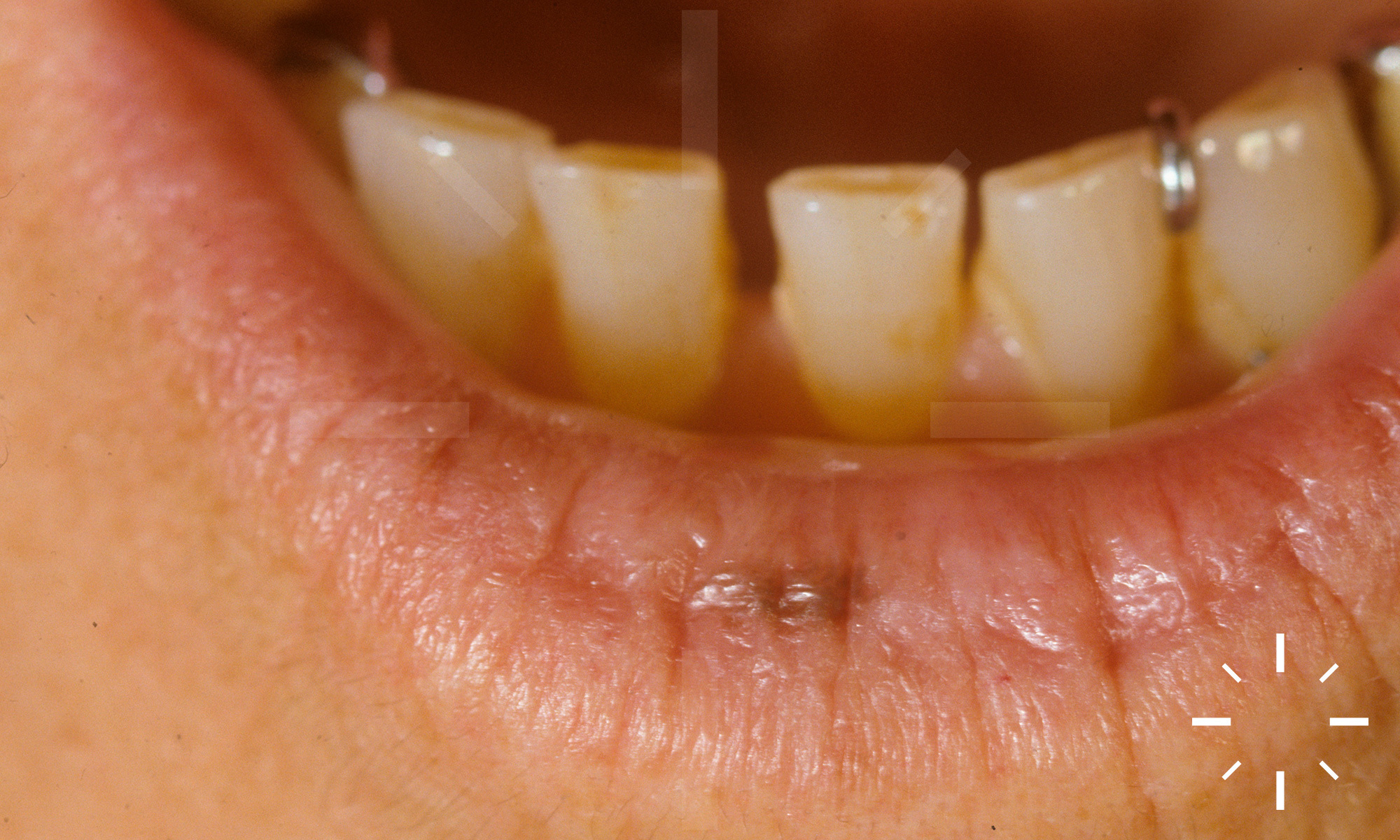
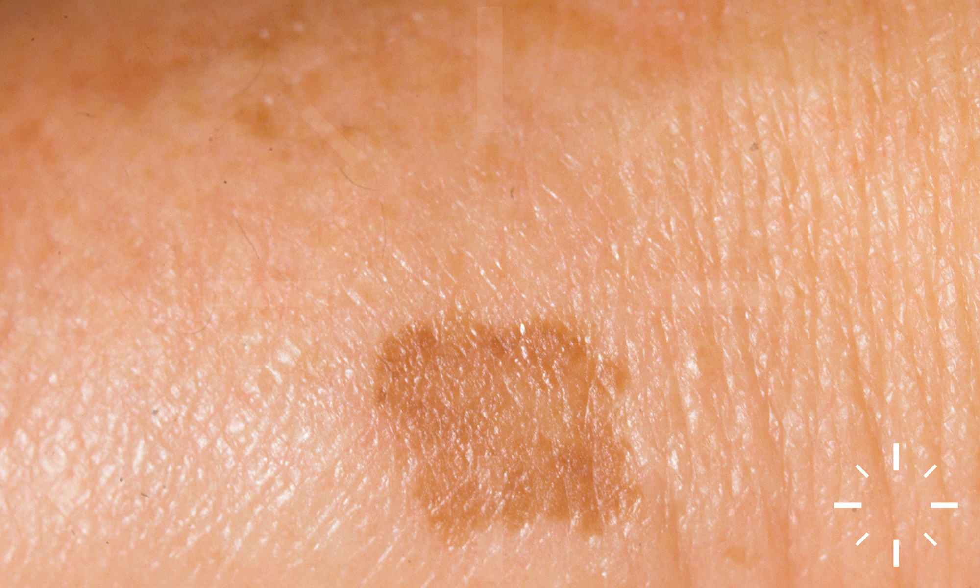
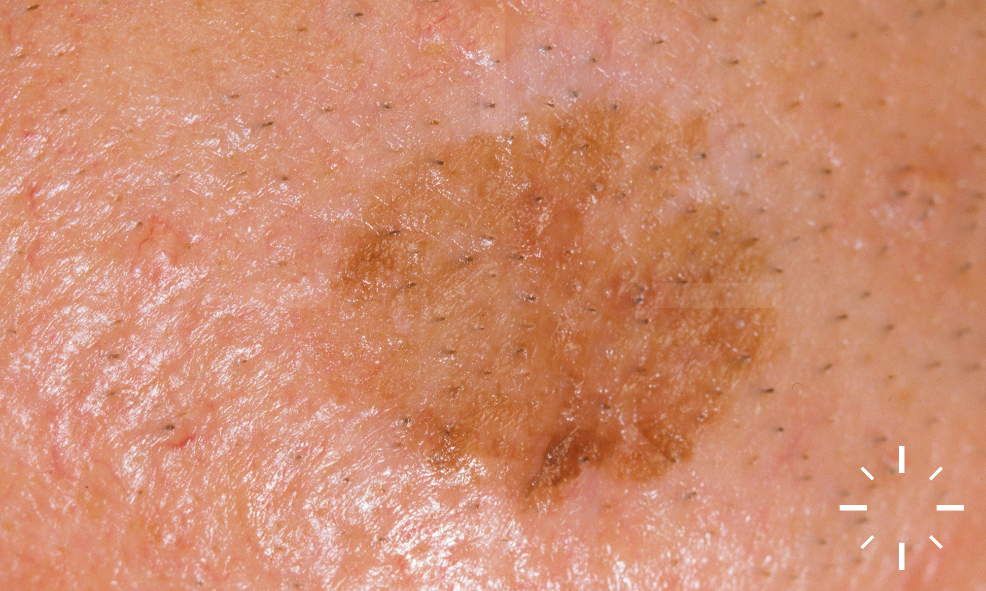
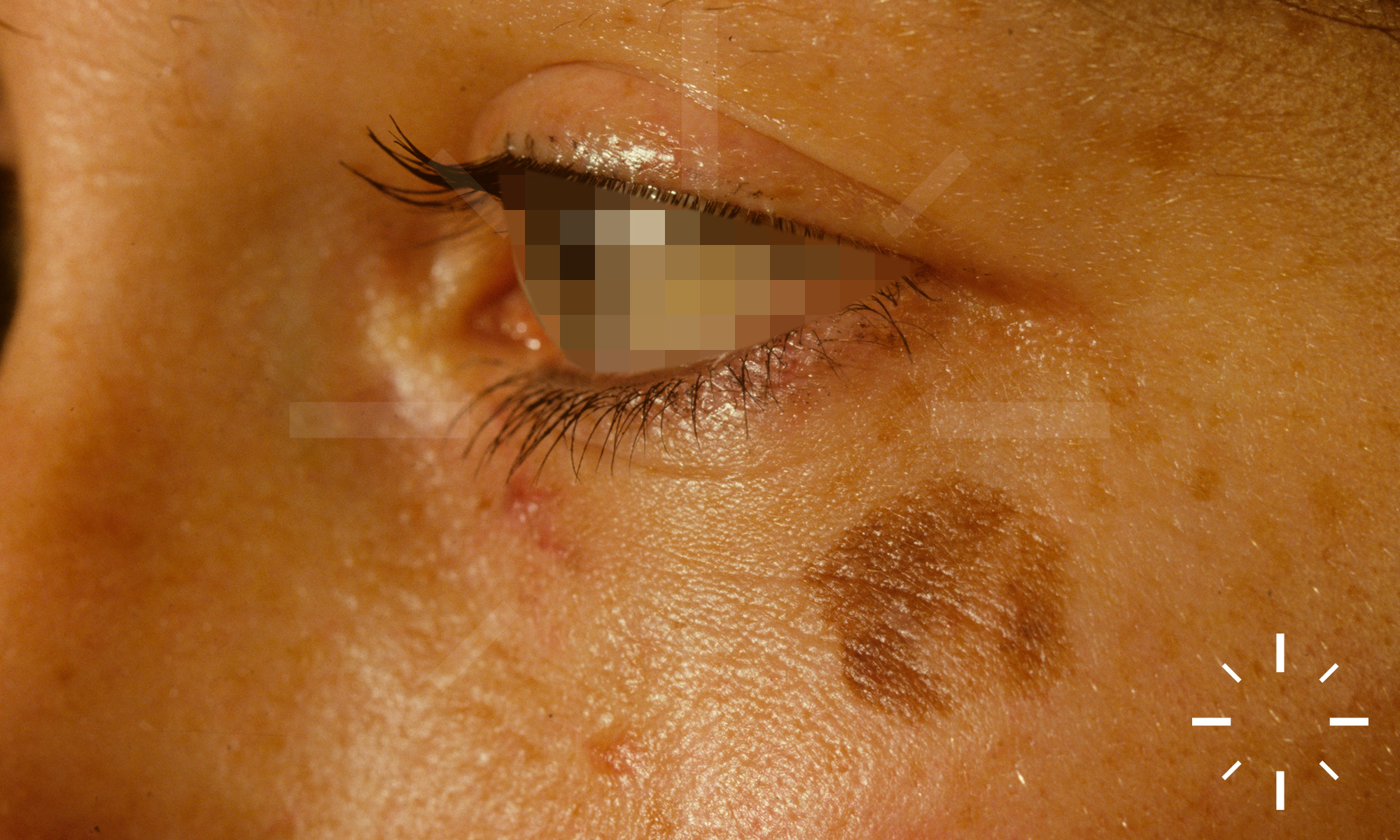
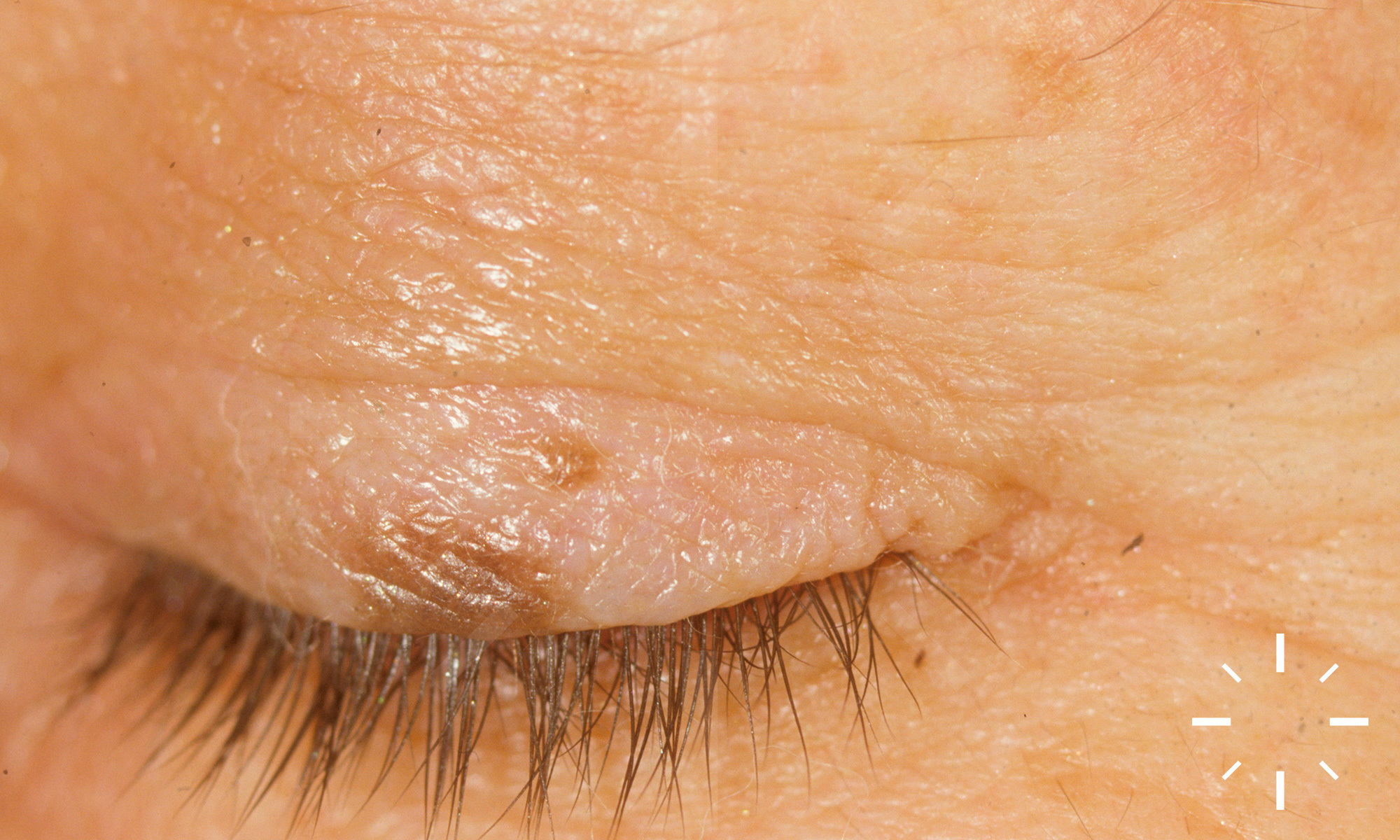
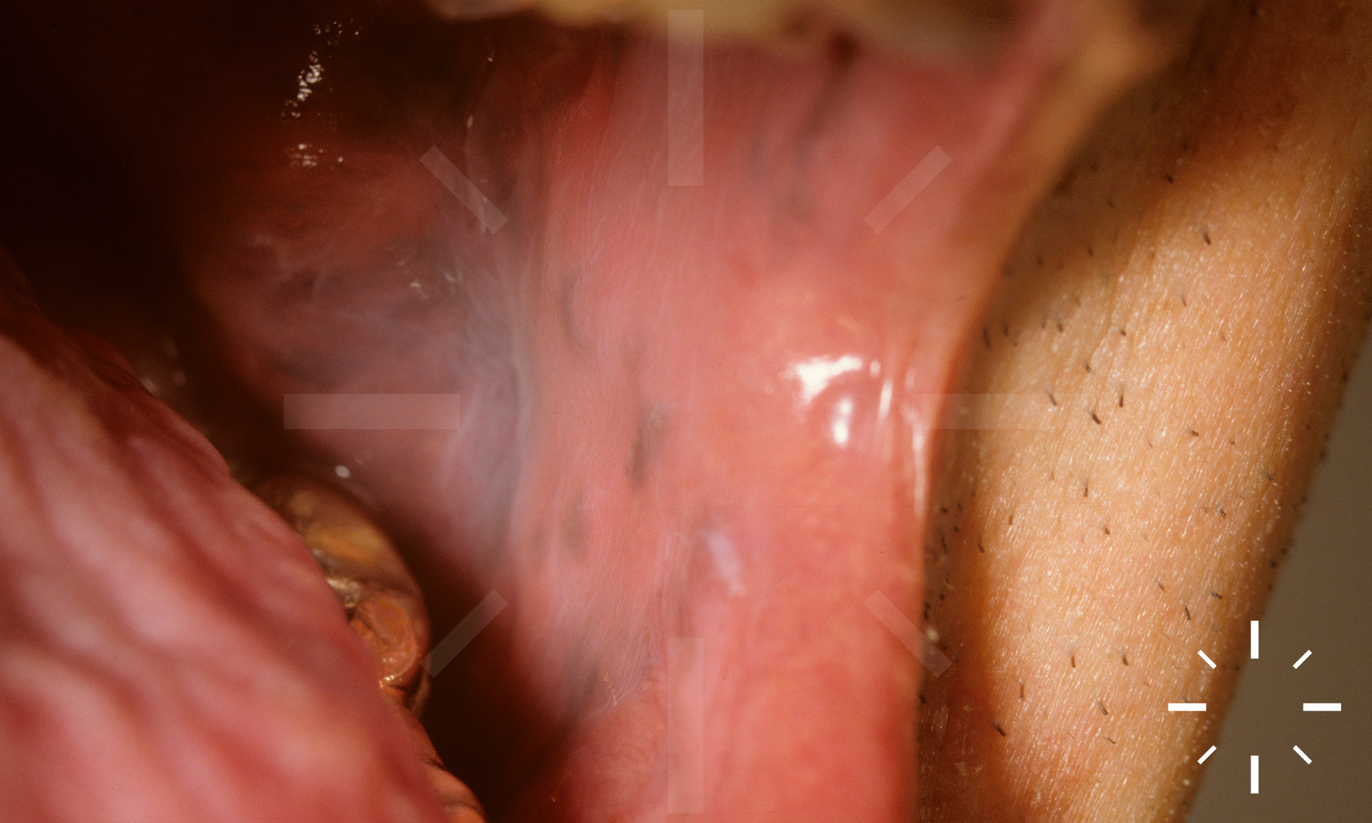
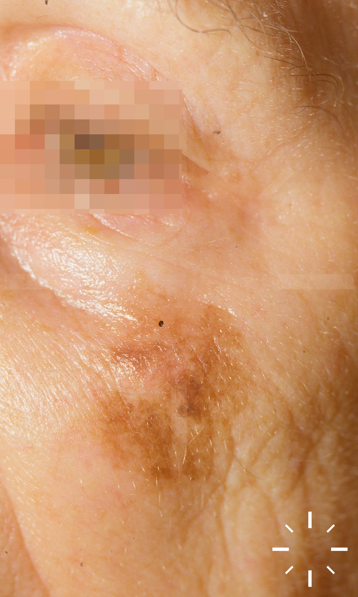
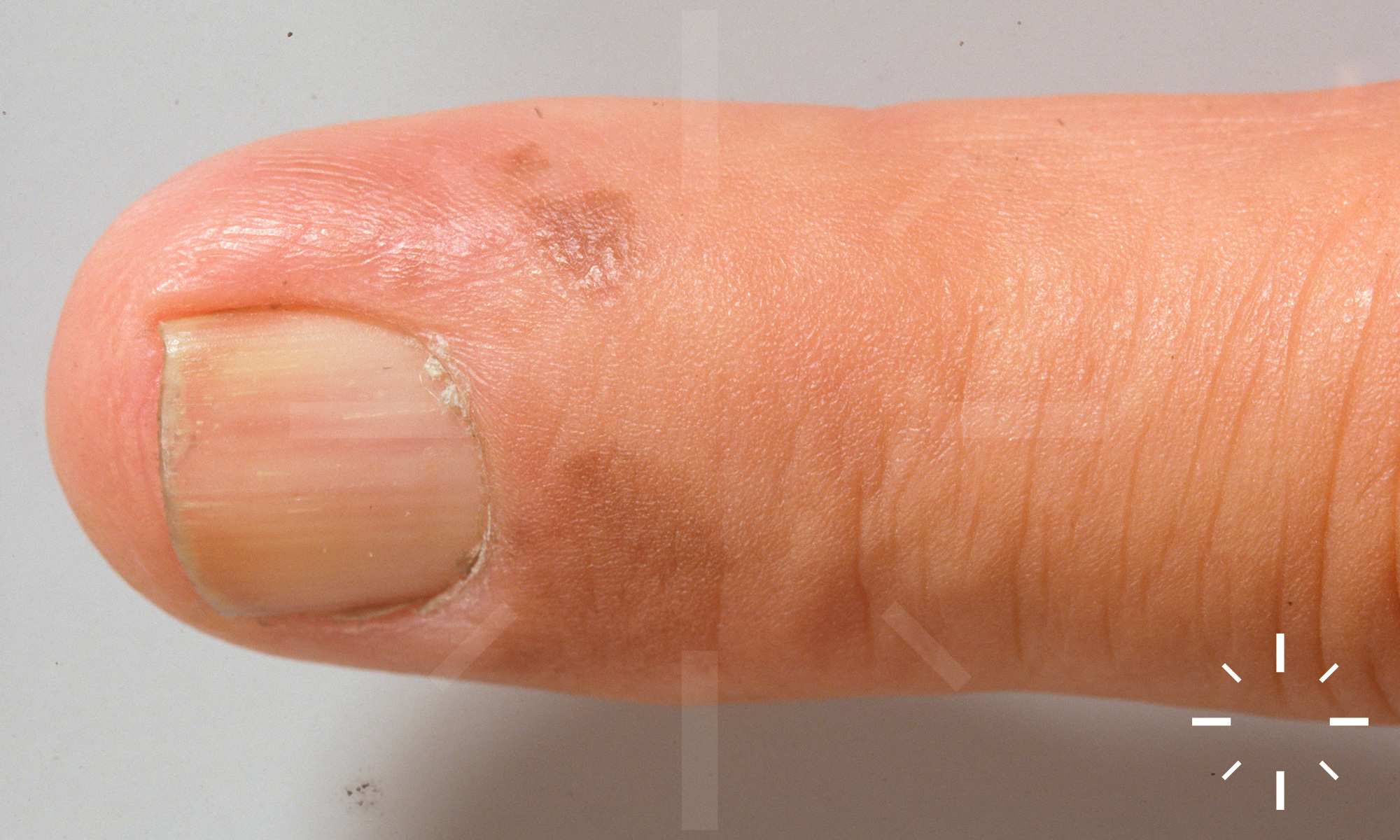
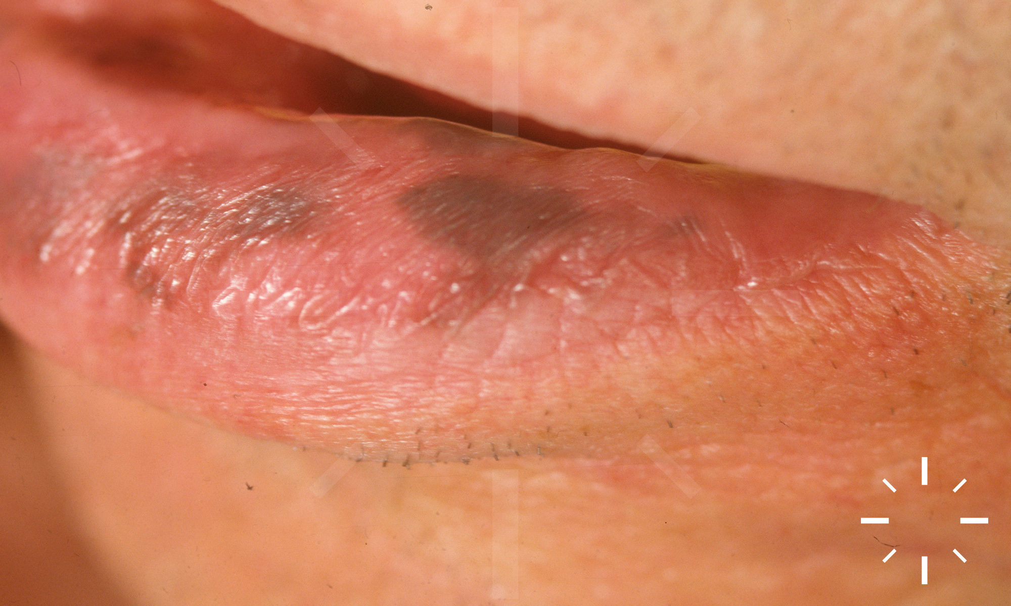
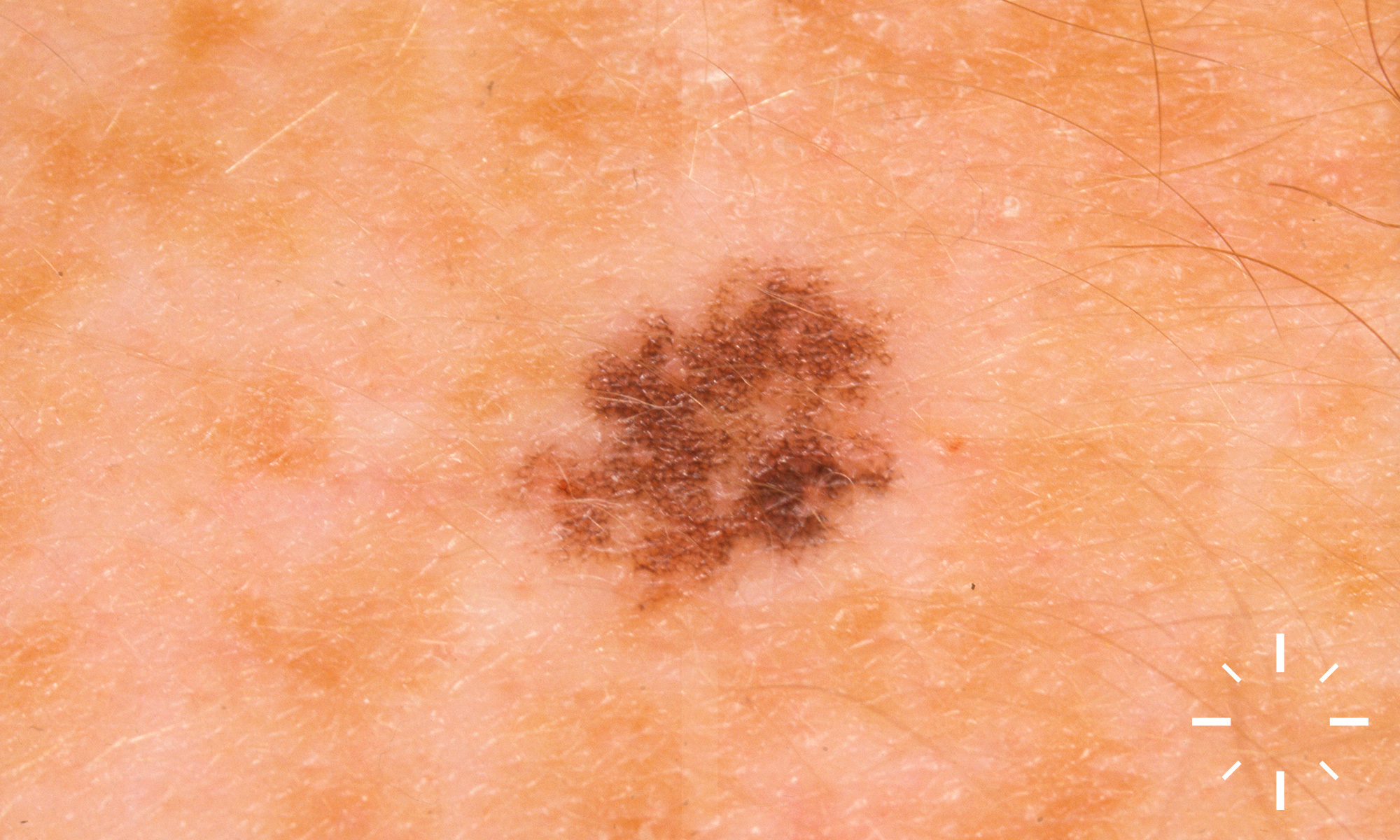
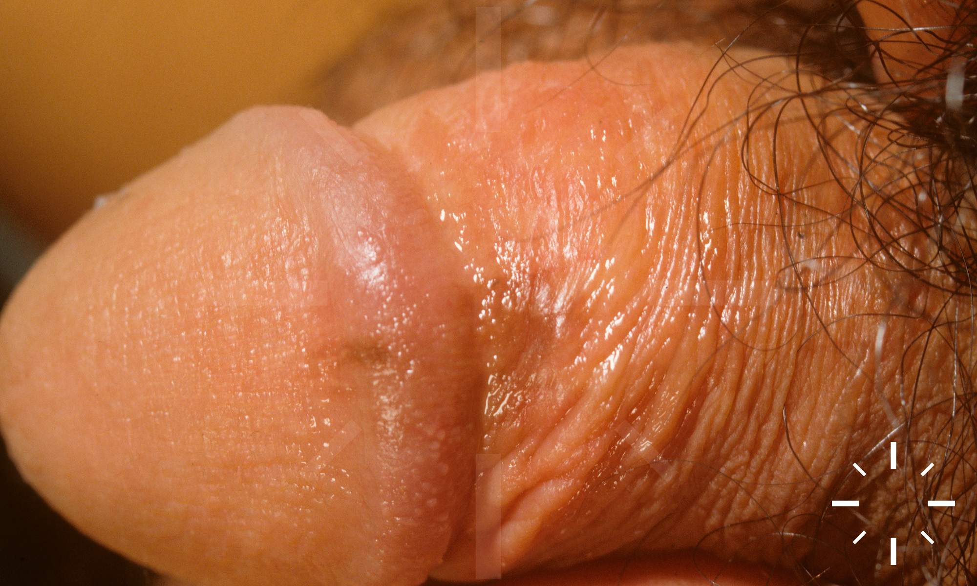
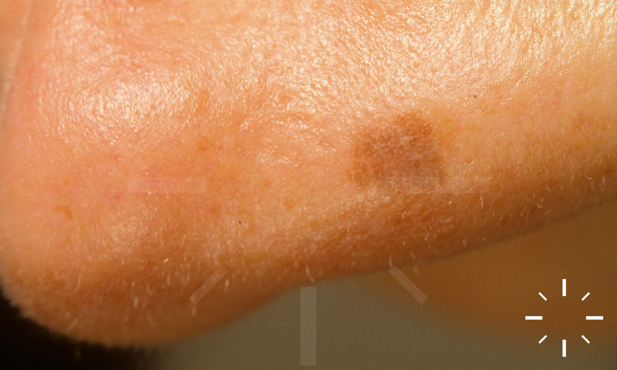
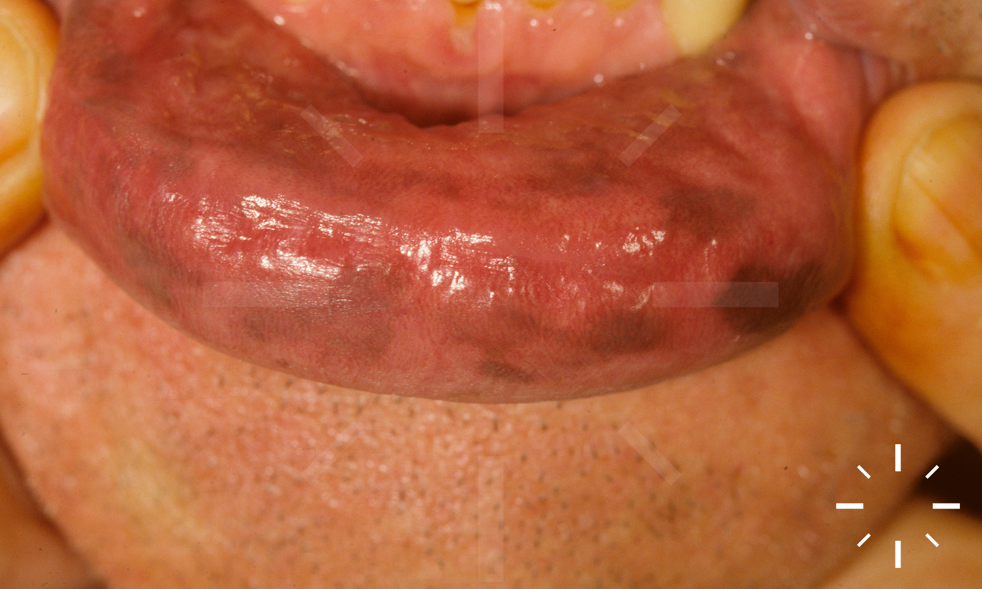
Lentigo simplex, Lentigo solaris

Last Updated: 2025-02-11

Author(s): Anzengruber F., Navarini A.

ICD11: 2F20.0Y

Lentigo juvenilis, lentigo benigna, lenticular stain, nevoid lentigo, naevus incipiens, lentigo of the mucous membrane, mucosal lentigo.

Cutaneous or mucous, acquired melanocyte hyperplasia, which appears as hyperpigmentation. It is UV-induced. There is no preference for light-exposed areas. The hyperpigmentation does not fade in winter.

  • Mostly occurring in the first years of life

  • In principle, however, lentigo simplex can occur at any age

  • Mostly phased progression

  • Lentigo simplex -> naevoid lentigo -> melanocytic naevus (junctional naevus to melanocytic naevus of the dermal type)

  • UV-induced lentigines can develop at any age

    • At an older age, the phased development decreases

  • People with skin type I according to Fitzpatrick and patients with peripheral neurofibromatosis or Peutz-Jeghers syndrome are less likely to develop the phased progression. 

    • This is the reason why redheads are prone to lentigines

  • Brownish, usually multiple, up to approx. 5 mm (but also larger up to 3.0 cm) in diameter, sharply defined hyperpigmentation with irregular pigmentation.

  • If lentigines occur in large numbers, they are referred to as lentiginosis.

  • Special form 

    • Ink spot lentigo or reticular lentigo 

      • Black, sometimes bizarrely shaped reticular lentigo often occurring after intensive UV exposure in the shoulder girdle area.


Avoid common mistakes:

  • Lentigines which occur mainly in red-haired children, are called ephelides

  • If perioral lentiginosis occurs, a gastroenterological presentation must be made to rule out Peutz-Jeghers syndrome

  • Clinic
  • Biopsy if necessary

  • Irregular, often club-shaped, retracted epidermal rete cones

  • Basal hyperpigmentation with increased number of individual melanocytes in the basal layer

  • Single dermal localised melanophages

  • No melanocyte nests

  • No solar elastosis

Rare transitions to malignant melanoma possible.

Sunscreen.

Often transition into a junctional nevus.

  1. Samuelov, L., et al, Extensive lentigo simplex, linear epidermolytic naevus and epidermolytic naevus comedonicus caused by a somatic mutation in KRT10. Br J Dermatol, 2015. 173(1): p. 293-6.
  2. Hafner, C., et al, The absence of BRAF, FGFR3, and PIK3CA mutations differentiates lentigo simplex from melanocytic nevus and solar lentigo. J Invest Dermatol, 2009. 129(11): p. 2730-5.
  3. Erkek, E., et al, Type I hereditary punctate keratoderma associated with widespread lentigo simplex and successfully treated with low-dose oral acitretin. Arch Dermatol, 2006. 142(8): p. 1076-7.
  4. Awaya, A., K. Watanabe, and S. Kato, Individuals exhibiting conspicuous nevi (lentigo simplex) are resistant to allergic rhinitis/conjunctivitis (pollinosis), but those who do not show increased susceptibility to pollinosis. Microbiol Immunol, 2003. 47(1): p. 101-3.
  5. McCarthy, D.J., Lentigo simplex. J Am Podiatr Med Assoc, 1987. 77(10): p. 539-43.
  6. Horikoshi, T., K. Jimbow, and S. Sugiyama, Comparison of macromelanosomes and autophagic giant melanosome complexes in nevocellular nevi, lentigo simplex and malignant melanoma. J Cutan Pathol, 1982. 9(5): p. 329-39.
  7. Hirone, T. and Y. Eryu, Ultrastructure of giant pigment granules in lentigo simplex. Acta Derm Venereol, 1978. 58(3): p. 223-9.