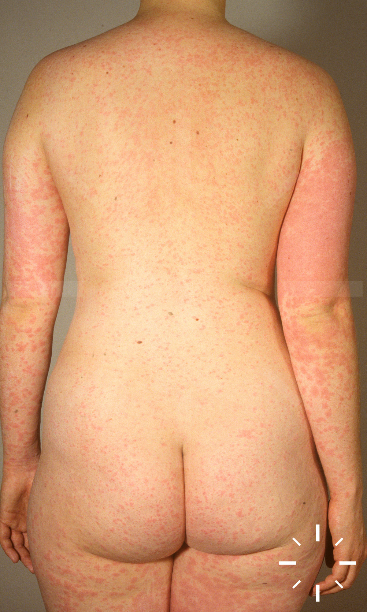
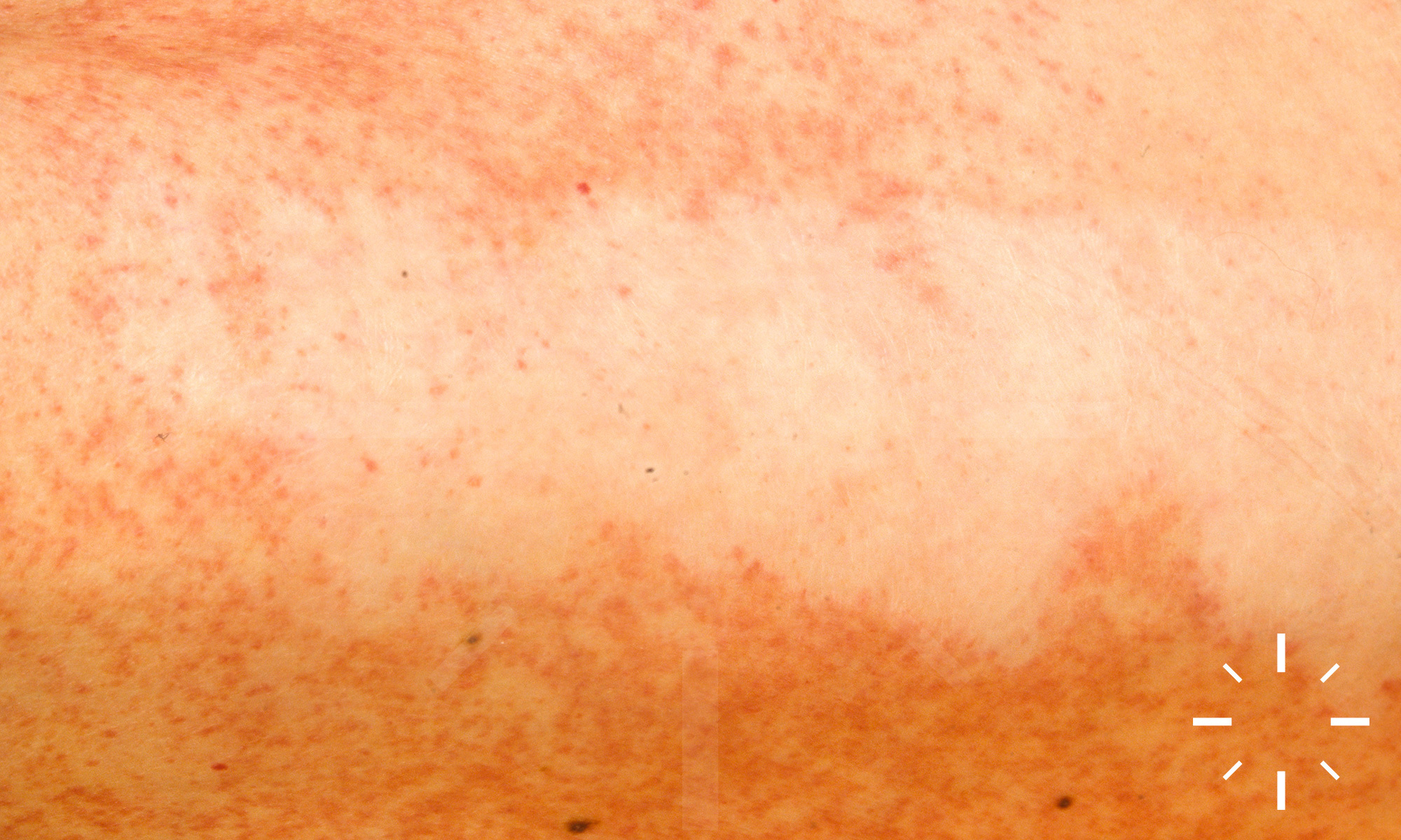
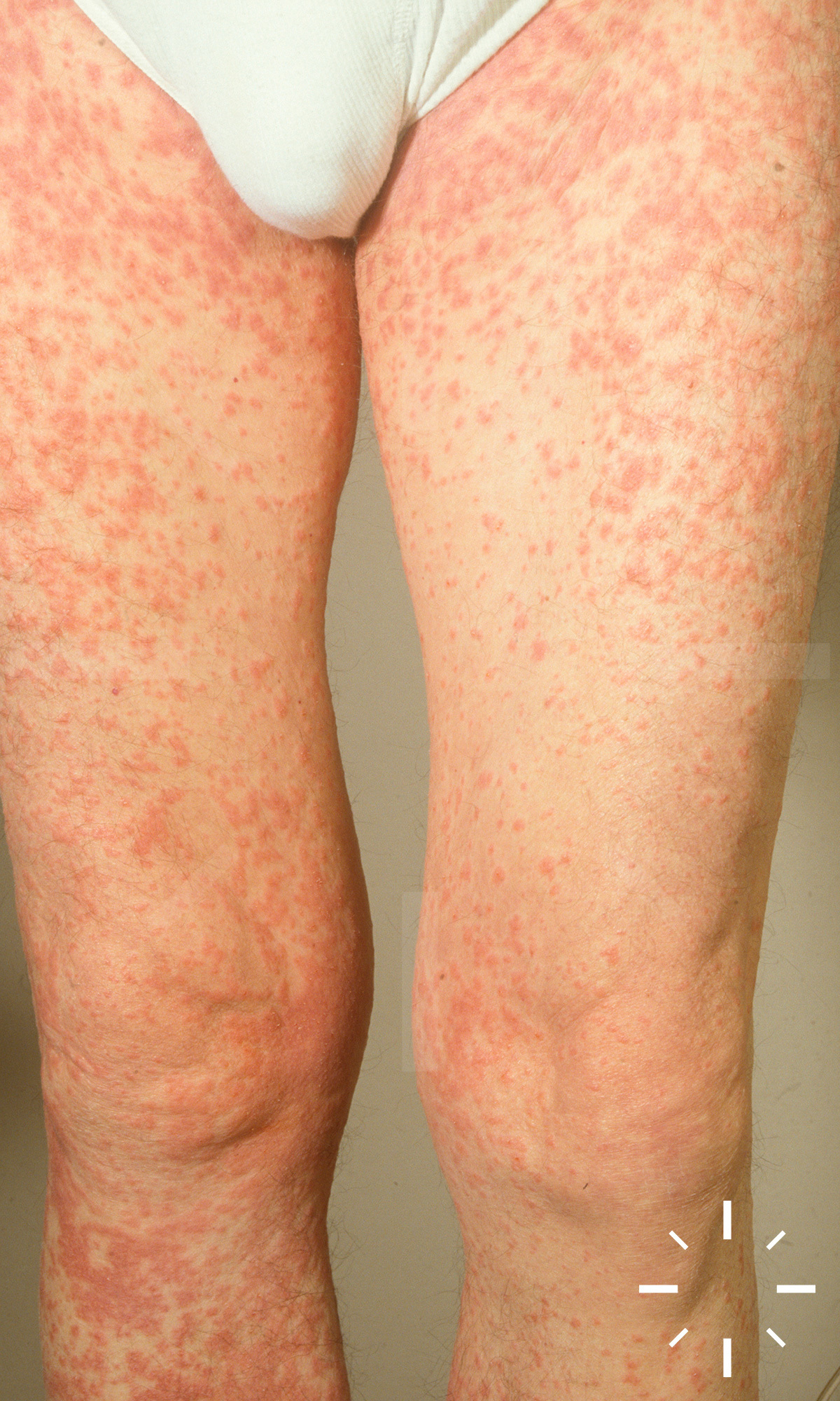
Stevens Johnson syndrome and toxic epidermal necrolysis
Last Updated: 2025-02-11
Author(s): Anzengruber F.
ICD11: EB13.2
- Staphylococcal scaled skin syndrome (SSSS)
- Erythema exsudativum multiforme
- Pemphigus vulgaris
- Pemphigus foliaceus
- Bullous pemphigoid
- Stevens Johnson syndrome: Stevens & Johnson, 1922
- Toxic epidermal necrolysis : Lyell, 1956
Stevens Johnson syndrome, SJS, toxic epidermal necrolysis, TEN, Lyell syndrome, epidermolysis necroticans combustiformis, epidermolysis acuta toxica
Seldom, serious, potentially lethal, mucocutaneous disease, which is usually triggered by taking medication. It leads to detachment of the epidermis and the appearance of necrosis. Stevens Johnson syndrome (SJS) and toxic epidermal necrolysis (TEN) correspond to one clinical picture. Cheilitis and stomatitis occur in 100% of cases. The area of spread to the rest of the skin is different.
-
Classification
-
Stevens Johnson syndrome: <10% of the body surface, mucous membranes are affected (eyes, oral cavity, genital).
-
SJS/TEN overlap syndrome: > 10% to < 30% of the body surface area, mucous membranes are affected (eyes, oral cavity, genital).
-
TEN: > 30% of the body surface area, mucous membranes are affected (eyes, oral cavity, genital).
-
- Incidence (for SJS, SJS/TEN and TEN): 2-7/1,000,000/per annum
- SJS:TEN = approx. 3:1
- 100-fold increased incidence in HIV-positive patients
- Women > men
- Frequently triggering medication: Mnemonic SATAN
- Sulfonamide antibiotics: Sulfamethoxazole (Bactrim), sulfasalazine
- Allopurinol
- Tetracyclines & other antibiotics: Amoxicillin, cephalosporins, quinolones, etc.
- Anticonvulsants: carbamazepine, lamotrigine, phenytoin, etc.
- NSAIDs
- Virostatics (HAART): nevirapine
- Infections
- Mycoplasma pneumoniae
- Cytomegalovirus infections
- Vaccinations
- Contrast media
- Food
- Bone marrow transplants
- Radiotherapy
- Predisposing factors
- HIV infection
- Genetics:
- HLA: HLA-B*1502, HLA-A*3101, B*5801, HLA-B*1502, HLA-A*3101, HLA-B*5801.
- Polymorphisms
- Prodromal stage:
- Deteriorated AZ, fever
- Influenza-like symptoms
- Facultative:
- Photophobia
- Conjunctival pruritus
- Pain when swallowing
- Myalgias
- Arthralgias
- Purulent rhinitis
- Joint complaints
- Generalised lymphadenopathy
- Liver and spleen involvement
- Facultative:
- Mucocutaneous symptoms
- Mostly appear after 1-3, but sometimes after up to 14 days
- Lesions usually start on the face or trunk before spreading to other parts of the body
- The capillitium is not affected, but palmoplantar involvement is also seen in some cases.
- The duration of mucocutaneous symptoms is reported to be 8-12 days.
- Mucosa:
- In 90% of cases, the oral mucosa is affected
- Erosions and ulcerations
- Haemorrhagic crusts on the lips.
- The eyes (conjunctivitis, sometimes purulent) are almost always affected
- Crusy lesions on the upper and lower eyelids.
- Corneal ulcer, panophthalmitis, photophobia, synechiae, symblepharon.
- Genital erosions
- Synechiae and stenoses can develop in the labia and urethra.
- Urethritis, urinary retention, cystitis
- Involvement of the pharynx can be observed in almost all patients
- Rarely, the trachea, bronchi, oesophagus and intestines may also be involved.
- In 90% of cases, the oral mucosa is affected
- Cutaneous manifestation:
- Epidermal detachments, erosions and partly ulcers
- Cocardiform plaques and maculae
- Often scarlatiniform, stem-accentuated exanthema
- Formation of vesicles and bullae
- Nikolski phenomenon I (blisters can be triggered on healthy skin or non-blistered skin can be displaced when lateral pressure is applied)
- Nikolski phenomenon II (existing blisters can be displaced by lateral pressure)
- Asboe-Hansen sign (the blister expands laterally in response to pressure
- Coarse lamellar desquamation/the skin peels off over a large area
- Necroses
- Onycholysis, if applicable
- Reepithelialisation occurs after a few days
- Laboratory
- Increased ESR, CRP, leukocytosis
- Egg electrophoresis (increased α- & γ-globulin fraction)
- Anaemia (approx. 15%)
- Lymphopenia (frequent)
- Neutropenia
- Occurs in approx. 1/3 of all patients
- Often masked by systemic glucocorticoids
- Correlated with a poor prognosis
- Eosinophilia (20%)
- Elevated transaminases (approx. 15%)
- Proteinuria and haematuria (5%)
- Bacteraemia (approx. 27%)
- Signs of a severe course:
- Urea >10 mmol/L
- Glucose >14 mmol/L
- ALT (up to 2-3 times the value)
- Anamnesis (medication anamnesis)
- Clinical picture
- Biopsy
- Rapid dermatopathological section! Important to differentiate from SSSS. Therefore has a pronounced therapeutic consequence!
- Direct immunofluorescence
- With the "algorithm of drug causality for epidermal necrolysis" (ALDEN)
- Laboratory
-
Laboratory daily: BB, small coagulation status, chemogram incl. CRP, urea
and bicarbonate, blood cultures if required, serum electrophoresis
-
Initial one-off: IgA quantitative in serum and HIV if necessary
-
- Bact. Swabs from skin and mucous membranes, blood cultures, cultures from urinary catheters and PEG probes should be repeated every 2 days if necessary!
- SCORTEN: Assess the severity using the SCORTEN (severity-of-illness) score* (if the score is >2, organise a burns IPS transfer (USZ)). Repeat daily
- Differentiation from: EEMM, SSSS, bullous autoimmune dermatoses, other severe drug reactions
- SJS/TEN: Disseminated, generalised, mucous membrane
-
Initial macular (dolent!), target lesions, blisters, "scalded skin"
-
Can affect internal organs: Pneumonitis, gastrointestinal tract
-
Fever, reduction in general condition
-
Lymphopenia
-
Histology: Epidermal necrosis
-
Begin after taking medication in case of new sensitisation: 1 - 4 weeks without pre-sensitisation
-
- AGEP: Disseminated, generalised, initially possibly flexural. Erythroderma, "pinprick pustules", possibly with confluence and fine lamellar desquamation
- Can also affect internal organs (especially hepatopathy, nephropathy)
- Frequently fever > 39°C. Neutrophilia
- Histological subcorneal pustules.
- Start after taking medication in case of new sensitisation: 5 - 7 days without sensitisation, 1 -2 days with sensitisation (1 day for antibiotics)
- DHS/DRESS: Generalised, centrofacial swelling
-
Polymorphic exanthema including isolated pustules and blisters possible, rare courses without exanthema
-
Hepatopathy, pneumonitis, lymphadenopathy, nephritis, cardiopathy.
-
Fever, reduction in general condition
-
Eosinophilia, atypical lymphocytes, cytopenias
-
Histological lymphocyte infiltrates
-
Begin after taking medication in the case of new sensitisation: 2 - 6 weeks without pre-sensitisation
-
-
GBFDE (generalised bullous fixed drug exanthema): Generalised, mucosal, asymmetrically distributed
-
Livid maculae and plaques, blisters, intact skin in between
-
Internal organs not affected, slight fever possible. Normal blood count.
-
Histology epidermal necrosis
-
Begin after taking medication in case of new sensitisation: 5 - 7 days without sensitisation, 1 -2 days with sensitisation (1 day for antibiotics)
-
Subepidermal blister fissure, necrotic keratinocytes in the epidermis at the blister roof, otherwise unchanged str. corneum, ↑ eosinophils in the epidermis, spiny cells with ballooned degeneration. Lymphohistiocytic infiltrate in the corium, mainly arranged perivascularly.
- Hypoalbuminaemia
- Electrolyte imbalances
- Hypovolaemic shock
- Insulin resistance
- Catabolic metabolic state
- Multi-organ failure
- Bacterial infections
- Septic shock (frequently S. aureus and P. aeruginosa)
- Pneumonia or interstitial pneumonitis
- Necrosis of the oesophagus, small intestine and colon with perforation
- There are usually painful erosions in the area of the mucous membrane
- Estimation of the body surface infestation:
- 1 palm of the patient corresponds to approximately 1% of the body surface area in children and adults
- Rule of nines: rule for measuring body surface area
- Head ≙ 9% of the body surface area
- 1 arm ≙ 9% of the body surface area
- Breast & stomach ≙ 9% & 9% (= 18%) of the body surface
- 1 leg ≙ 18% (!) of the body surface area
- Genital ≙ 1% of the body surface
- SCORTEN (severity of illness score for toxic epidermal necrolysis)
- Scale designed to predict the probability of mortality
- Mortality (for SJS, SJS/TEN and TEN): approx. 30%
- Mortality (for SJS): 10%
- Mortality (TEN): >30%
SCORTEN (severity of illness score for toxic epidermal necrolysis) according to Bastuji-Garin | ||
---|---|---|
Age | ≥ 40 years | 1 |
Existing neoplasia? | Yes | 1 |
Body surface area affected | >10% | 1 |
Heart rate | ≥120/minute | 1 |
Serum urea | >10 mmol/L | 1 |
Serum glucose | >14 mmol/L | 1 |
Serum bicarbonate | >20 mmol/L | 1 |
Â
General measures
- Discontinue or replace suspected or non-essential medication immediately!
- Interdisciplinary management (intensive care, infection, derma, ophtha)
-
Large-scale hospitalisation on the IPS, certainly from SCORTEN >2 pts.
-
Isolation of the patient!
-
Special bed for decubitus prophylaxis
-
High-calorie diet for catabolic metabolic conditions, possibly tube or intravenous feeding
-
Nutrition
-
Liquid and electrolyte management
-
Pain therapy preferably with opiates (e.g. tramadol, morphine)
-
Targeted antibiotic treatment of infections
Â
Local therapy (daily dermatological ward round with wound expert - products available on the dermatological ward):
-
Blisters and erosions: Local antiseptic (e.g. Octenisept® Lsg or Prontosan
Woundgel® (X) or Polyhexanid-Crème® 0.04 %) Note: No sulphadiazine-silver (Flammazine® Crème or Ialugen plus® Crème) in case of sulphonamide-induced SJS/TEN. Leave blister covers in place.
-
Non-adherent film (e.g. Metalline® sheet (large surface area) or Mepitel® (small surface area))
-
Intact, reddened skin: Possibly topical corticosteroids (e.g. Elocom® cream, Locoid Crelo® emulsion) - Caution: No evidence, risk of infection!
-
Open full blisters aseptically if necessary, leave blister cover in place
-
Mucous membrane: Antiseptic (e.g. Dentohexin®-Lsg)
-
Lips: Externals containing dexpanthenol (e.g. Bepanthen® cream or ointment)
-
Eyes: According to ophthalmology! (Symblepharon prophylaxis)
Â
Â
For medium and heavy moulds:
General measures
- Treatment like burns
- Necroses should be debrided
- If > 30% of the KOF Ã intensive care
- Isolation for infection protection
- Lock
- Protective gown
- Mouth protection
- Gloves
- Decubitus prophylaxis
- Positioning on metal inlay
- Adequate fluid intake/balancing
- A patient with SJS or TEN needs 1/3 less fluid intake than burn patients
- Prophylactic antibiotics are not recommended!
- Increase the room temperature
- Optimally up to 32°C to reduce calorie consumption
- Nutrition
- If possible, passaged food
- Alternative: transnasal gastric tube
- Should be placed early
- Careful placement of the tube!
- Parenteral nutrition if necessary
- Thrombosis prophylaxis
- Support stockings
- Anticoagulation
- Low-molecular-weight heparin, for example
- Enoxaparin sodium 20 mg (low risk of thrombosis) or 40 mg (higher risk of thrombosis)
- Unfractionated heparin
- Ophthalmological consultation if necessary
- Gynaecological consultation
- Aim: avoidance of synechiae, stenoses
- Some authors recommend the intravaginal application of dilators to prevent stenosis and synechiae (Milex vaginal dilators). However, this must be clarified with the patient and we have no experience of this. Consult a gynaecologist
- Suppression of menstruation (with medication) to avoid crusts/synechiae
Â
Systemic therapy
- Ciclosporin p.o. 3-5 mg/kg bw daily
- Ciclosporin (Sandimmun® (i.v.), Sandimmun Neoral® (p.o.))
- First-line therapy for TEN, the RegiSCAR study was able to show ↑ survival rate
- Early administration after consultation with OA/LA/CA
- Dose 3 - 6 mg/kg bw/d (p.o. or i.v. according to ICU)
- Ciclosporin blood levels must be checked, trough level in the morning before drug administration, target 150 - 200 ng/ml
- Adverse drug reactions: uncontrolled art. Hypertension, hyperkalaemia, leucopenia, renal dysfunction, increased risk of infections, gastrointestinal disorders, headaches, etc.
- Add methylprednisolone 50 - 250 mg/d i.v. if necessary (analogue to GvHD 1-3 days)
- Systemic glucocorticoids
- Controversially discussed. There are studies that show a benefit, as well as studies that have demonstrated an increased complication rate and mortality
- In the RegiSCAR study, the overall benefit could not be verified
- The EuroSCAR study showed a mortality rate of 18% with the administration of systemic glucocorticoids
- All in all, therapy is not recommended
- If the decision is made in favour of treatment with systemic glucocorticoids, treatment should be started as soon as possible (preferably 24-48 hours after the first symptoms appear)
- Systemic glucocorticoids
- Prednisolone p.o. 1.0 mg/kg/d
- Methylprednisolone p.o. 40 mg 1x daily
- Intravenous immunoglobulins
- Second-line therapy for TEN, only if contraindicated for ciclosporin
- Administration i.v. 250-400 mg/kg bw daily over 3-5 days every 3-4 weeks
-
Early administration after consultation with OA/LA/CA
-
Dose 1 g/kg bw/d i.v. over 3 days according to the scheme (www.spitalpharmazie-
basel.ch, application information Privigen)
-
Contraindications: IgA deficiency (determine immunoglobulins in serum,
Do not delay administration by waiting for the results)
-
Restrictions on use: Renal insufficiency or risk factors for
Kidney failure (diabetes mellitus, volume depletion, sepsis, paraproteinaemia, >65y, nephrotoxic medication), heart failure, increased risk of thromboembolism
- Data are controversial
- The EuroSCAR study showed a mortality rate of 25% with the administration of IVIG
- Combination therapy with systemic glucocorticoids and IVIG
- Multiple retrospective analyses
- Data situation and dosage unclear, possible benefit
Â
More off-label alternatives
- Etanercept s.c. 50 mg once
- Infliximab i.v. 5mg kg bw once
- Wirz E., Scherer K., Häusermann P. Dermatology USB Standards (internal document, partly verbatim), 09 November 2017
- Arvalo JM, Lorente JA, Gonzalez-Herrada C , Jimenez-Reyes J. Treatment of Toxic Epidermal Necrolysis with Cyclosporin A. The Journal of Trauma: Injury, Infection, and Critical Care 2000;48:473-8.
- Assier H. Erythema Multiforme With Mucous Membrane Involvement and Stevens-Johnson Syndrome Are Clinically Different Disorders-Reply. Arch Dermatol 1996;132:711.
- Auquier-Dunant A, Mockenhaupt M, Naldi L, Correia O, Schröder W , Roujeau J-C. Correlations Between Clinical Patterns and Causes of Erythema Multiforme Majus, Stevens-Johnson Syndrome, and Toxic Epidermal Necrolysis. Arch Dermatol 2002;138.
- Bachot N, Revuz J , Roujeau J-C. Intravenous Immunoglobulin Treatment for Stevens-Johnson Syndrome and Toxic Epidermal Necrolysis. Arch Dermatol 2003;139.
- Bastuji-Garin S. Clinical Classification of Cases of Toxic Epidermal Necrolysis, Stevens-Johnson Syndrome, and Erythema Multiforme. Arch Dermatol 1993;129:92.
- Brown KM, Silver GM, Halerz M, Walaszek P, Sandroni A , Gamelli RL. Toxic Epidermal Necrolysis: Does Immunoglobulin Make a Difference? Journal of Burn Care & Rehabilitation 2004;25:81-8.
- Chung W-H, Hung S-I, Yang J-Y, Su S-C, Huang S-P, Wei C-Y et al. Granulysin is a key mediator for disseminated keratinocyte death in Stevens-Johnson syndrome and toxic epidermal necrolysis. Nature Medicine 2008;14:1343-50.
- Côté B. Clinicopathologic Correlation in Erythema Multiforme and Stevens-Johnson Syndrome. Arch Dermatol 1995;131:1268.
- Dietrich A, Kawakubo Y, Rzany B, Mockenhaupt M, Simon JC , Schopf E. Low N-acetylating capacity in patients with Stevens-Johnson syndrome and toxic epidermal necrolysis*. Experimental Dermatology 1995;4:313-6.
- Elias PM , Levy SW. Bullous impetigo. Occurrence of localised scalded skin syndrome in an adult. Arch Dermatol 1976;112:856-8.
- Fagot J-P, Mockenhaupt M, Bouwes-Bavinck J-N, Naldi L, Viboud C , Roujeau J-C. Nevirapine and the risk of Stevens-Johnson syndrome or toxic epidermal necrolysis. AIDS 2001;15:1843-8.
- Fouchard N, Bertocchi M, Roujeau J-C, Revuz J, Wolkenstein P , Bastuji-Garin S. SCORTEN: A Severity-of-Illness Score for Toxic Epidermal Necrolysis. Journal of Investigative Dermatology 2000;115:149-53.
- Garcia-Doval I, LeCleach L, Bocquet H, Otero X-L , Roujeau J-C. Toxic epidermal necrolysis and Stevens-Johnson syndrome. Arch Dermatol 2000;136.
- Gubinelli E, Canzona F, Tonanzi T, Raskovic D , Didona B. Toxic epidermal necrolysis successfully treated with etanercept. The Journal of Dermatology 2009;36:150-3.
- Halebian PH, Corder VJ, Madden MR, Finklestein JL , Shires GT. Improved Burn Centre Survival of Patients with Toxic Epidermal Necrolysis Managed without Corticosteroids. Annals of Surgery 1986;204:503-12.
- Halevy S, Ghislain P-D, Mockenhaupt M, Fagot J-P, Bouwes Bavinck JN, Sidoroff A et al. Allopurinol is the most common cause of Stevens-Johnson syndrome and toxic epidermal necrolysis in Europe and Israel. Journal of the American Academy of Dermatology 2008;58:25-32.
- Hebert AA , Bogle MA. Intravenous immunoglobulin prophylaxis for recurrent Stevens-Johnson syndrome. Journal of the American Academy of Dermatology 2004;50:286-8.
- Hess Schmid M , Elsner P. Unusual haemorrhagic variant of Stevens-Johnson syndrome in an HIV-infected patient. Der Hautarzt 1999;50:52-5.
- Hewitt J , Ormerod AD. Toxic epidermal necrolysis treated with cyclosporine. Clin Exp Dermatol 1992;17:264-5.
- Hunger RE, Hunziker T, Buettiker U, Braathen LR , Yawalkar N. Rapid resolution of toxic epidermal necrolysis with anti-TNF-α treatment. Journal of Allergy and Clinical Immunology 2005;116:923-4.
- Kamanabroo D. Plasmapheresis in Severe Drug-Induced Toxic Epidermal Necrolysis. Arch Dermatol 1985;121:1548.
- Kardaun SH , Jonkman MF. Dexamethasone Pulse Therapy for Stevens-Johnson Syndrome/Toxic Epidermal Necrolysis. Acta Derm Venereol 2007;87:144-8.
- Laffitte E, Nenadov Beck M, Hofer M, Hohl D , Panizzon RG. Severe Stevens-Johnson syndrome induced by contrast medium iopentol (ImagopaqueR). Br J Dermatol 2004;150:376-8.
- Leckie MJ. Stevens-Johnson syndrome in association with hydroxychloroquine treatment for rheumatoid arthritis. Rheumatology 2002;41:473-4.
- Lonjou C, Borot N, Sekula P, Ledger N, Thomas L, Halevy S et al. A European study of HLA-B in Stevens???Johnson syndrome and toxic epidermal necrolysis related to five high-risk drugs. Pharmacogenetics and Genomics 2008;18:99-107.
- Lyell A. TOXIC EPIDERMAL NECROLYSIS: AN ERUPTION RESEMBLING SCALDING OF THE SKIN. Br J Dermatol 1956;68:355-61.
- Mockenhaupt M, Viboud C, Dunant A, Naldi L, Halevy S, Bavinck JNB et al. Stevens-Johnson Syndrome and Toxic Epidermal Necrolysis: Assessment of Medication Risks with Emphasis on Recently Marketed Drugs. The EuroSCAR Study. Journal of Investigative Dermatology 2008;128:35-44.
- Nassif A, Bensussan A, Bachot N, Bagot M, Boumsell L, Roujeau J-C et al. Drug Specific Cytotoxic T-Cells in the Skin Lesions of a Patient with Toxic Epidermal Necrolysis. Journal of Investigative Dermatology 2002;118:728-33.
- Ng CY, Yeh YT, Wang CW, Hung SI, Yang CH, Chang YC et al. Impact of the HLA-B58:01 Allele and Renal Impairment on Allopurinol-Induced Cutaneous Adverse Reactions. J Invest Dermatol 2016.
- Paul C, Wolkenstein P, Adle H, Wechsler J, Garchon HJ, Revuz J et al. Apoptosis as a mechanism of keratinocyte death in toxic epidermal necrolysis. Br J Dermatol 1996;134:710-4.
- Pereira FA, Mudgil AV , Rosmarin DM. Toxic epidermal necrolysis. Journal of the American Academy of Dermatology 2007;56:181-200.
- Prins C. Treatment of Toxic Epidermal Necrolysis With High-Dose Intravenous Immunoglobulins. Arch Dermatol 2003;139:26.
- Redondo P, Felipe I, Pena A, Aramendia JM , Vanaclocha V. Drug-induced hypersensitivity syndrome and toxic epidermal necrolysis. Treatment with N-acetylcysteine. Br J Dermatol 1997;136:645-6.
- Revuz J , Roujeau JC. [Lyell's staphylococcal syndrome or bullous impetigo?] Nouv Presse Med 1980;9:1097.
- Revuz JE , Roujeau JC. Advances in toxic epidermal necrolysis. Seminars in Cutaneous Medicine and Surgery 1996;15:258-66.
- Roujeau J-C. Genetic Susceptibility to Toxic Epidermal Necrolysis. Arch Dermatol 1987;123:1171.
- Roujeau J-C. Toxic epidermal necrolysis (Lyell syndrome). Arch Dermatol 1990;126:37.
- Roujeau J-C, Chosidow O, Saiag P , Guillaume J-C. Toxic epidermal necrolysis (Lyell syndrome). Journal of the American Academy of Dermatology 1990;23:1039-58.
- Roujeau J-C, Kelly JP, Naldi L, Rzany B, Stern RS, Anderson T et al. Medication Use and the Risk of Stevens-Johnson Syndrome or Toxic Epidermal Necrolysis. New England Journal of Medicine 1995;333:1600-8.
- Rzany B, Hering O, Mockenhaupt M, Schroder W, Goerttler E, Ring J et al. Histopathological and epidemiological characteristics of patients with erythema exudativum multiforme major, Stevens-Johnson syndrome and toxic epidermal necrolysis. Br J Dermatol 1996;135:6-11.
- Rzany B, Mockenhaupt M, Baur S, Schröder W, Stocker U, Mueller J et al. Epidemiology of erythema exsudativum multiforme majus, Stevens-Johnson syndrome, and toxic epidermal necrolysis in Germany (1990-1992): Structure and results of a population-based registry. Journal of Clinical Epidemiology 1996;49:769-73.
- Saiag P, Caumes E, Chosidow O, Revuz J , Roujeau J-C. Drug-induced toxic epidermal necrolysis (Lyell syndrome) in patients infected with the human immunodeficiency virus. Journal of the American Academy of Dermatology 1992;26:567-74.
- Sassolas B, Haddad C, Mockenhaupt M, Dunant A, Liss Y, Bork K et al. ALDEN, an algorithm for assessment of drug causality in Stevens-Johnson syndrome and toxic epidermal necrolysis: comparison with case-control analysis. Clin Pharmacol Ther 2010;88:60-8.
- Schneck J, Fagot J-P, Sekula P, Sassolas B, Roujeau JC , Mockenhaupt M. Effects of treatments on the mortality of Stevens-Johnson syndrome and toxic epidermal necrolysis: A retrospective study on patients included in the prospective EuroSCAR Study. Journal of the American Academy of Dermatology 2008;58:33-40.
- Schöpf E. Toxic Epidermal Necrolysis and Stevens-Johnson Syndrome. Arch Dermatol 1991;127:839.
- Shay E, Kheirkhah A, Liang L, Sheha H, Gregory DG , Tseng SCG. Amniotic Membrane Transplantation as a New Therapy for the Acute Ocular Manifestations of Stevens-Johnson Syndrome and Toxic Epidermal Necrolysis. Survey of Ophthalmology 2009;54:686-96.
- Stern RS. Improving the Outcome of Patients With Toxic Epidermal Necrolysis and Stevens-Johnson Syndrome. Arch Dermatol 2000;136.
- Ting W, Stone MS, Racila D, Scofield RH , Sontheimer RD. Toxic epidermal necrolysis-like acute cutaneous lupus erythematosus and the spectrum of the acute syndrome of apoptotic pan-epidermolysis (ASAP): a case report, concept review and proposal for new classification of lupus erythematosus vesiculobullous skin lesions. Lupus 2004;13:941-50.
- Tougeron-Brousseau B, Delcampe A, Gueudry J, Vera L, Doan S, Hoang-Xuan T et al. Vision-Related Function After Scleral Lens Fitting in Ocular Complications of Stevens-Johnson Syndrome and Toxic Epidermal Necrolysis. American Journal of Ophthalmology 2009;148:852-9.e2.
- Trent JT, Kirsner RS, Romanelli P , Kerdel FA. Analysis of Intravenous Immunoglobulin for the Treatment of Toxic Epidermal Necrolysis Using SCORTEN. Arch Dermatol 2003;139.
- Trent JT, Ma F, Kerdel F, Fien S, French LE, Romanelli P et al. Dose of intravenous immunoglobulin and patient survival in SJS and toxic epidermal necrolysis. Expert Review of Dermatology 2007;2:299-303.
- Watanabe H, Koide R , Iijima M. Toxic Epidermal Necrolysis Arising as a Sequela of Drug-induced Hypersensitivity Syndrome. Acta Dermato Venereologica 2012;92:214-5.
- Wetter DA, Davis MDP, Yiannias JA, Gibson LE, Dahl MV, El-Azhary RA et al. Effectiveness of Intravenous Immunoglobulin Therapy for Skin Disease Other Than Toxic Epidermal Necrolysis: A Retrospective Review of Mayo Clinic Experience. Mayo Clinic Proceedings 2005;80:41-7.
- Wolff K , Tappeiner G. Treatment of Toxic Epidermal Necrolysis. Arch Dermatol 2003;139.
- Wolkenstein P, Carri??re Vr, Charue D, Bastuji-Garin S, Revuz J, Roujeau J-C et al. A slow acetylator genotype is a risk factor for sulphonamide-induced toxic epidermal necrolysis and Stevens-Johnson syndrome. Pharmacogenetics 1995;5:255-8.
- Wolkenstein P, Chosidow O, Flechet M-L, Robbiola O, Paul M, Dume L et al. Patch testing in severe cutaneous adverse drug reactions, including Stevens-Johnson syndrome and toxic epidermal necrolysis. Contact Dermatitis 1996;35:234-6.
- Wolkenstein P, Latarjet J, Roujeau J-C, Duguet C, Boudeau S, Vaillant L et al. Randomised comparison of thalidomide versus placebo in toxic epidermal necrolysis. The Lancet 1998;352:1586-9.
- Wolkenstein PE, Roujeau JC , Revuz J. Drug-Induced Toxic Epidermal Necrolysis. Clinics in Dermatology 1998;16:399-408.
- Bastuji-Garin S et al. (2000) SCORTEN: a severity-of-illness score for toxic epidermal necrolysis. J Invest Dermatol 115: 149-153
- Nirken, M. (2016). Stevens-Johnson syndrome and toxic epidermal necrolysis: Pathogenesis, clinical manifestations, and diagnosis. Uptodate.com. Retrieved 22 May 2016, from http://www.uptodate.com/contents/stevens-johnson-syndrome-and-toxic-epidermal-necrolysis-pathogenesis-clinical-manifestations-and-diagnosis?source=search_result&search=stevens-johnson-syndrom&selectedTitle=1%7E150
- High, W. (2016). Stevens-Johnson syndrome and toxic epidermal necrolysis: Management, prognosis, and long-term sequelae. Uptodate.com. Retrieved 22 May 2016, from http://www.uptodate.com/contents/stevens-johnson-syndrome-andtoxic-epidermal-necrolysis-management-prognosis-and-long-term-sequelae?source=search_result&search=stevens-johnsonsyndrom&selectedTitle=2%7E150
