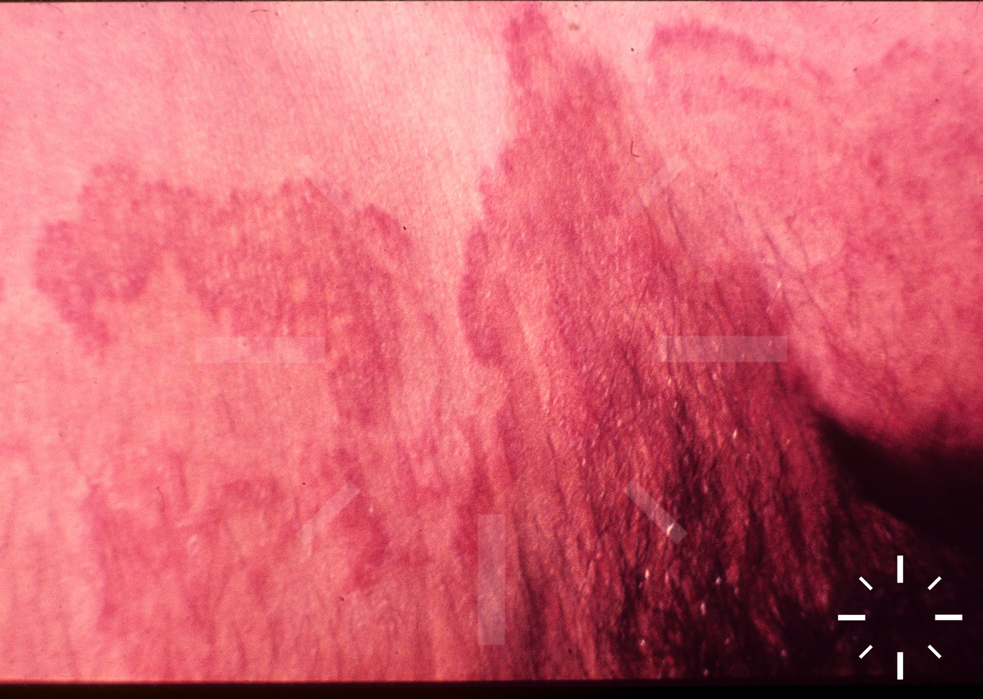
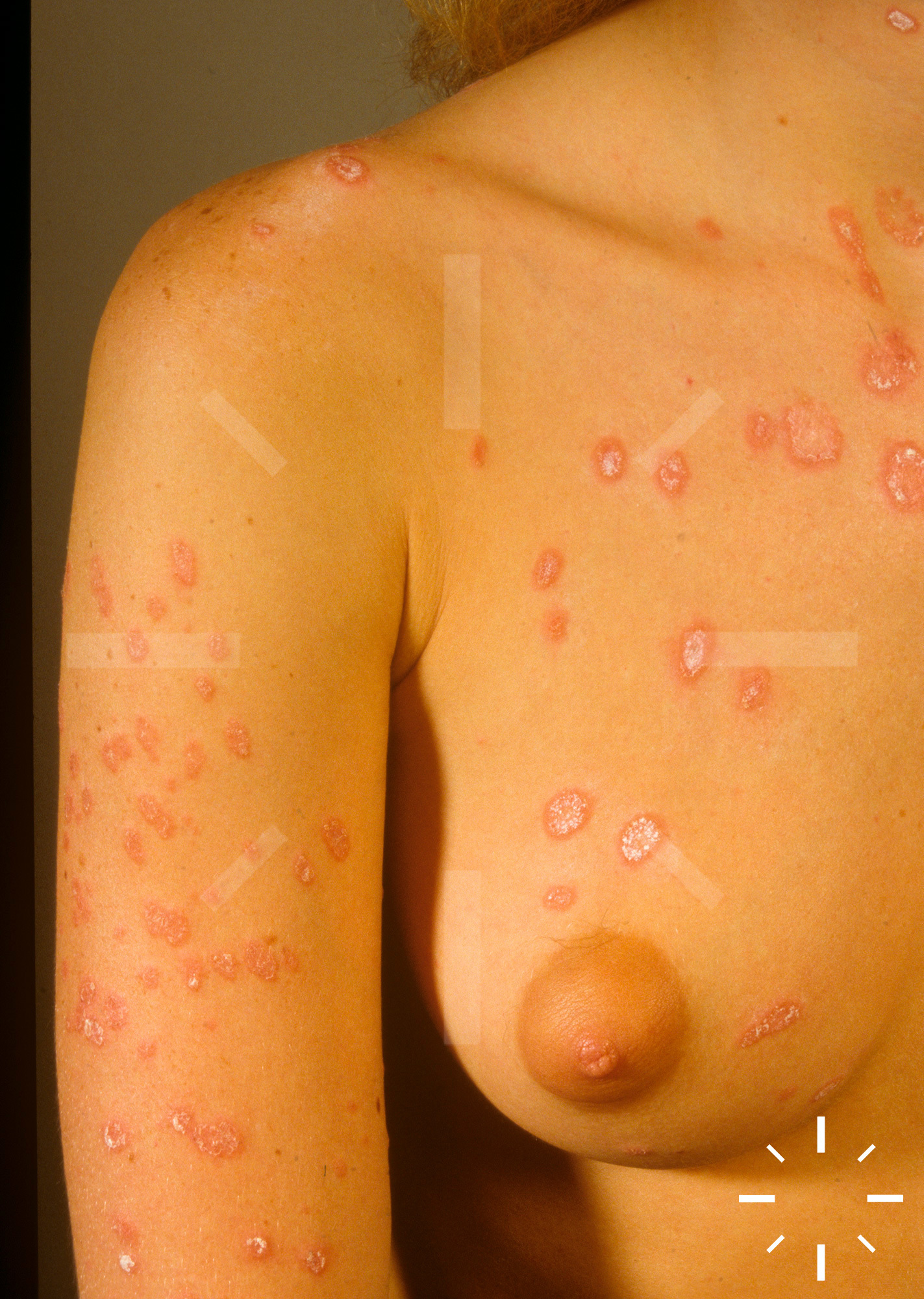
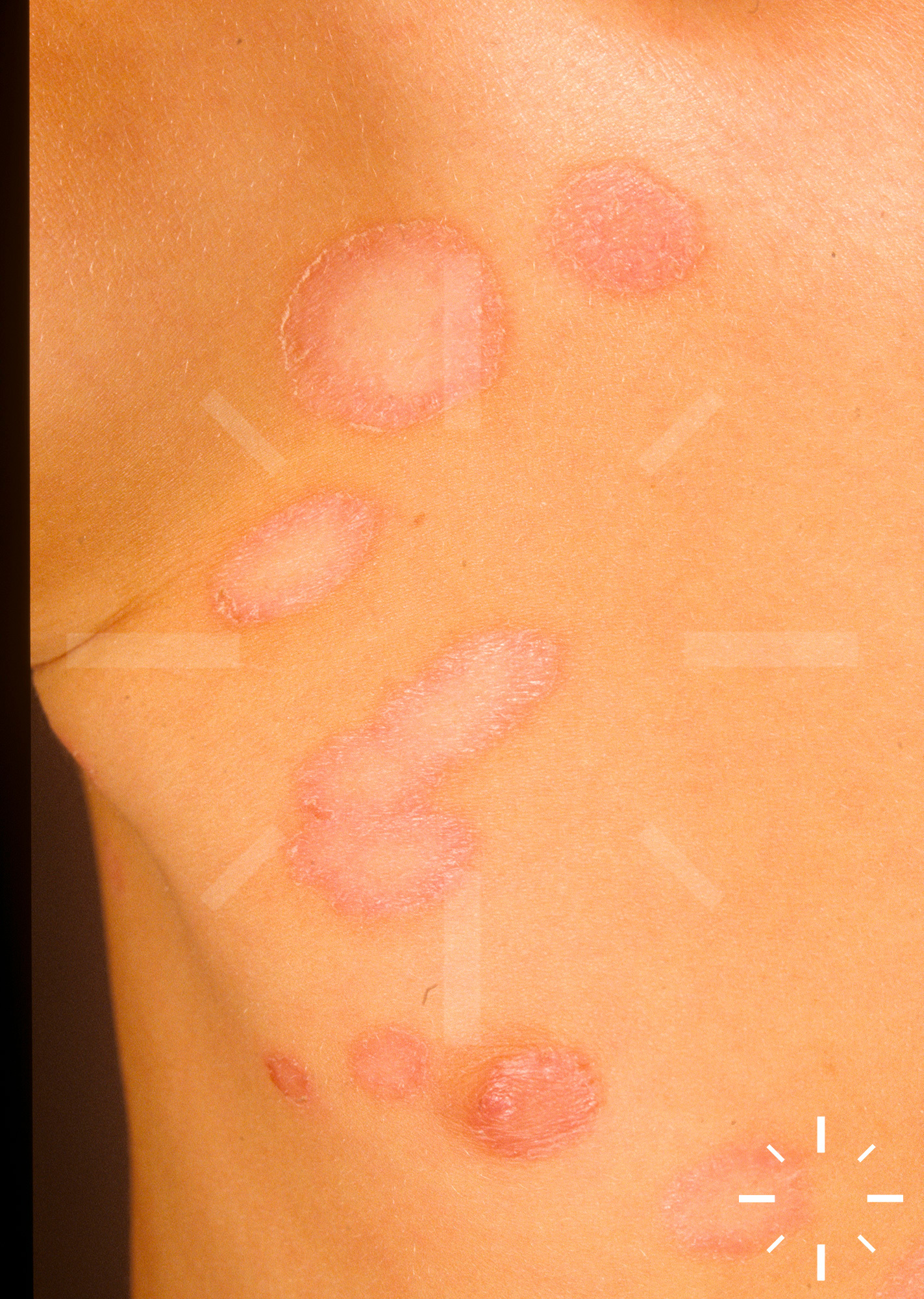
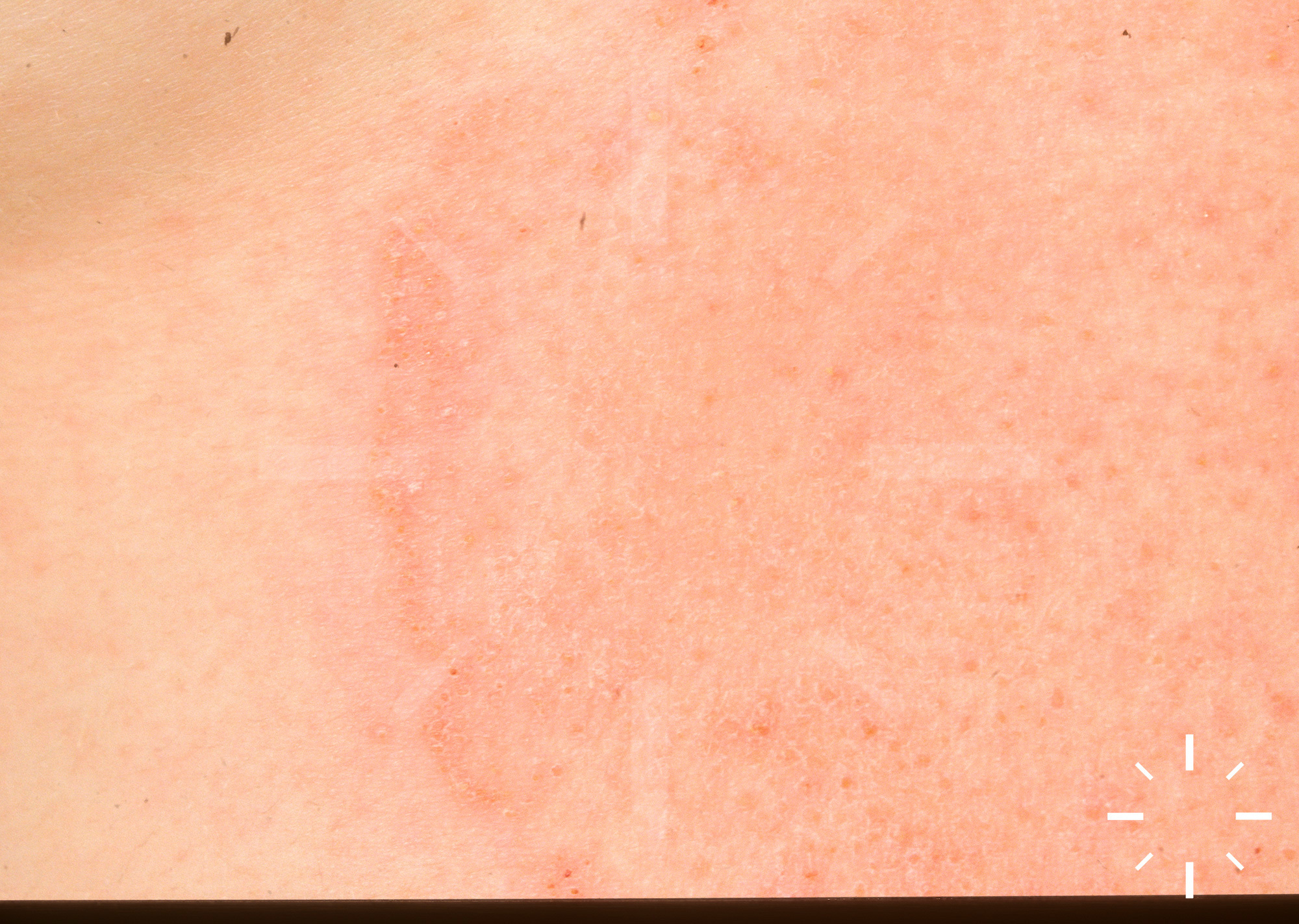
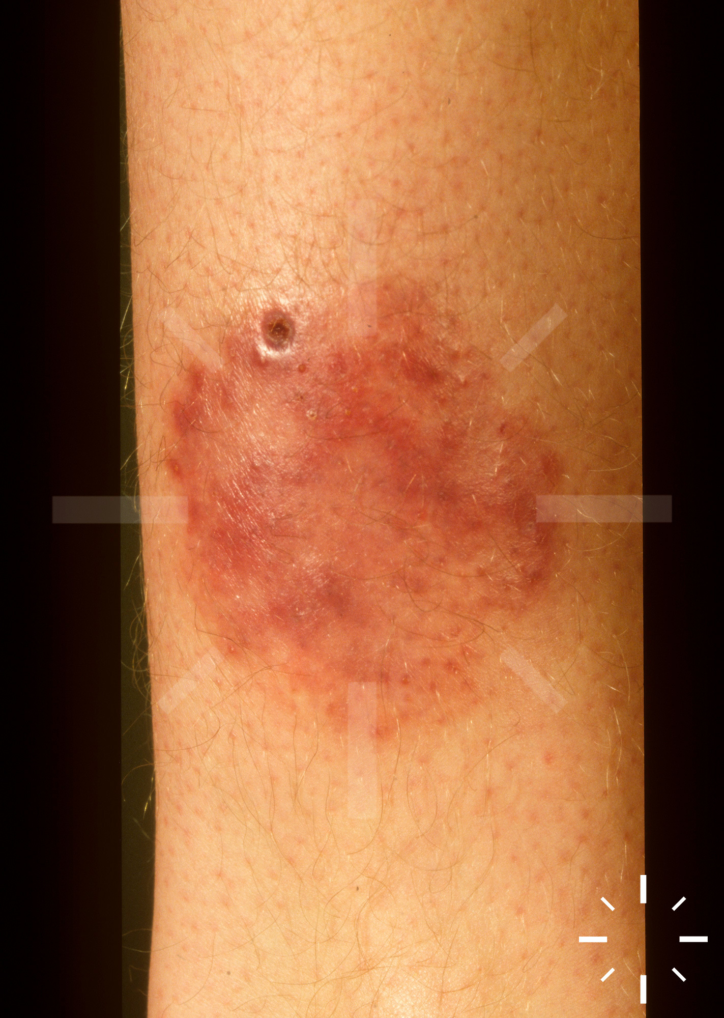
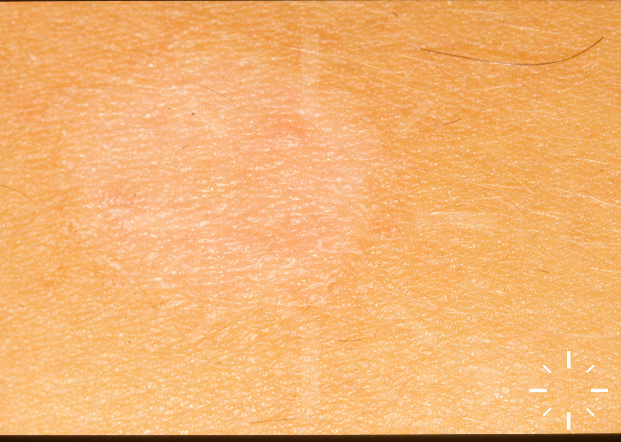
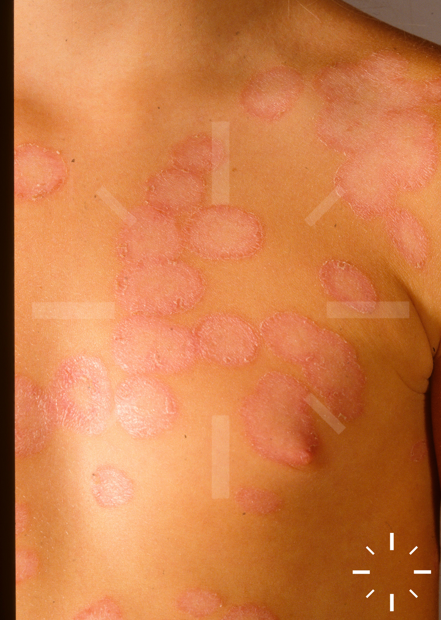
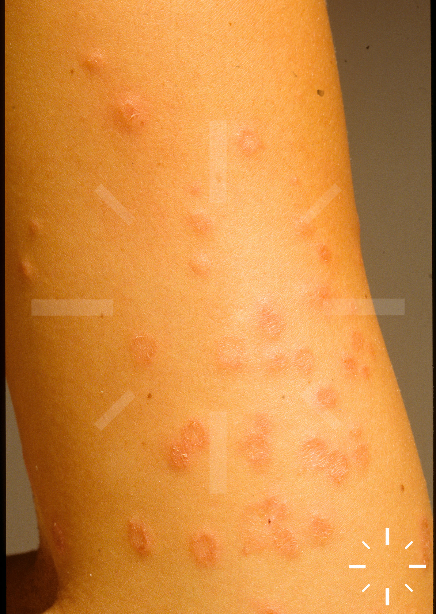
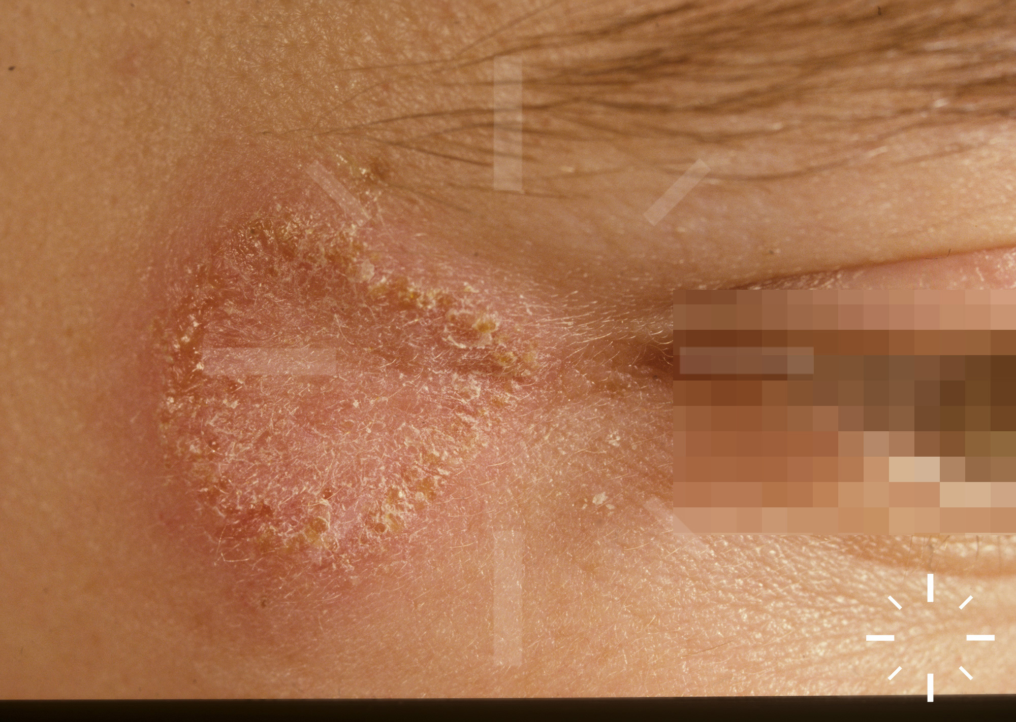
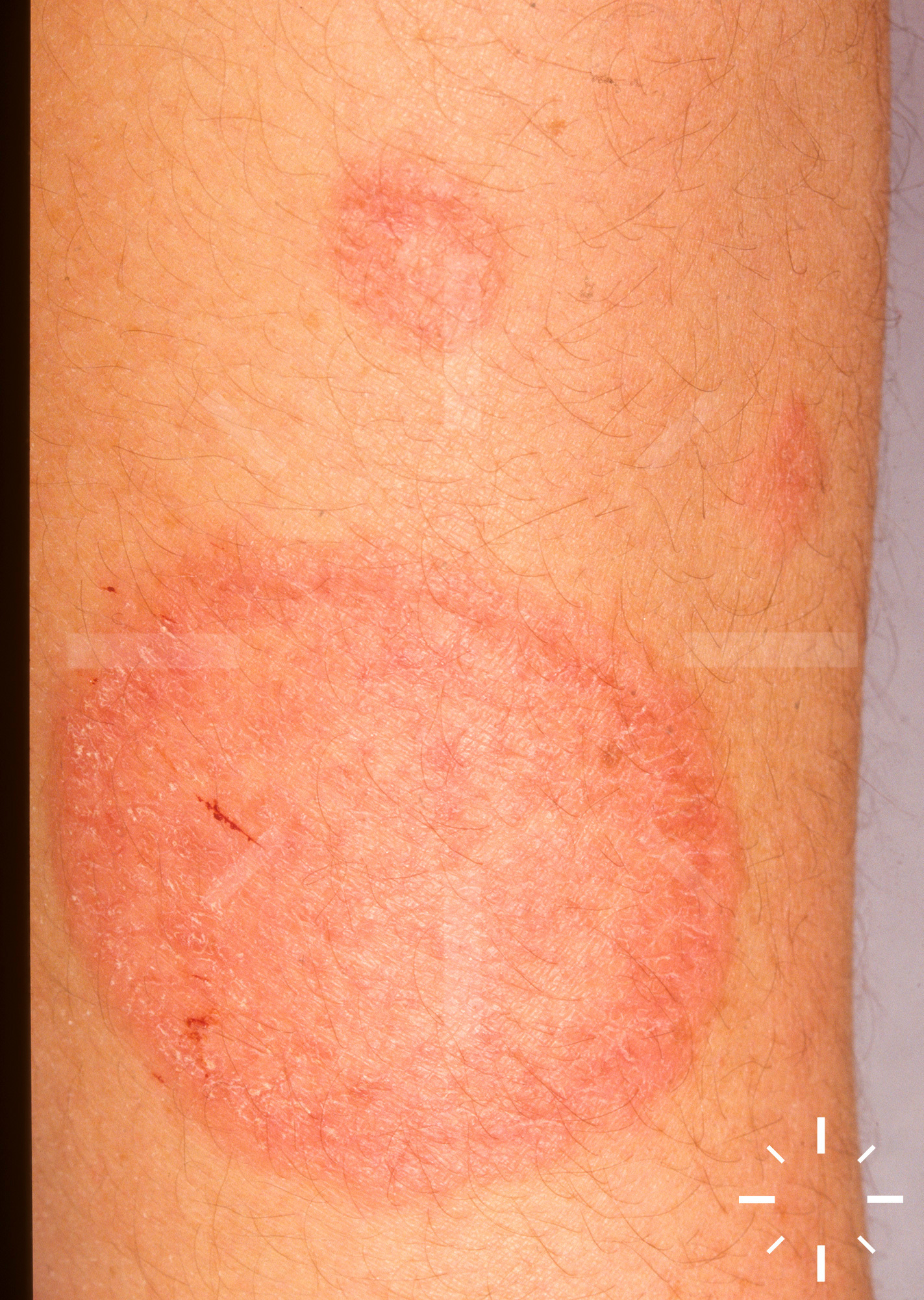
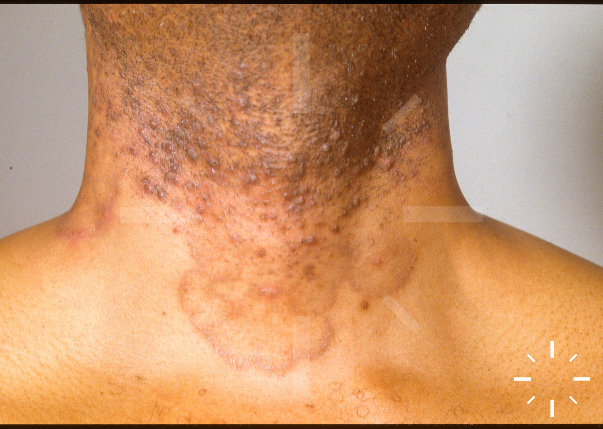
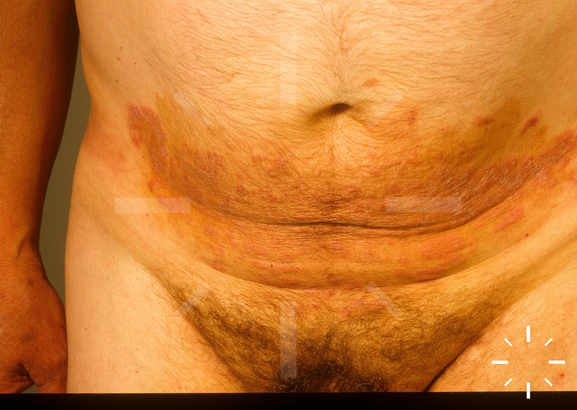
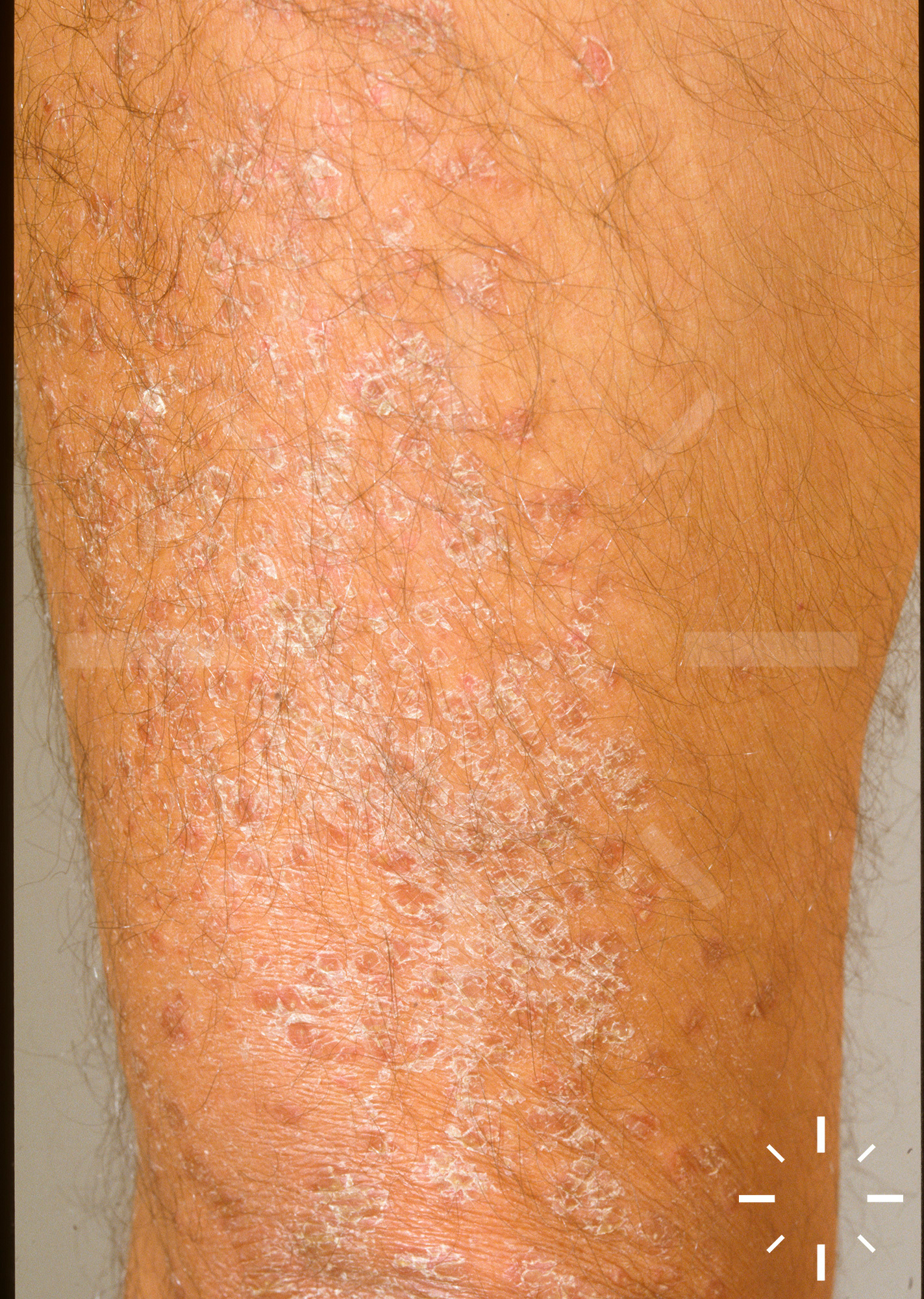
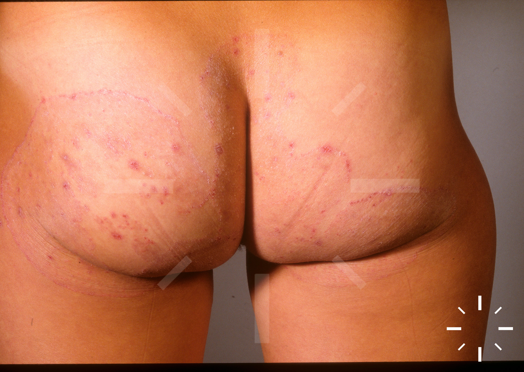
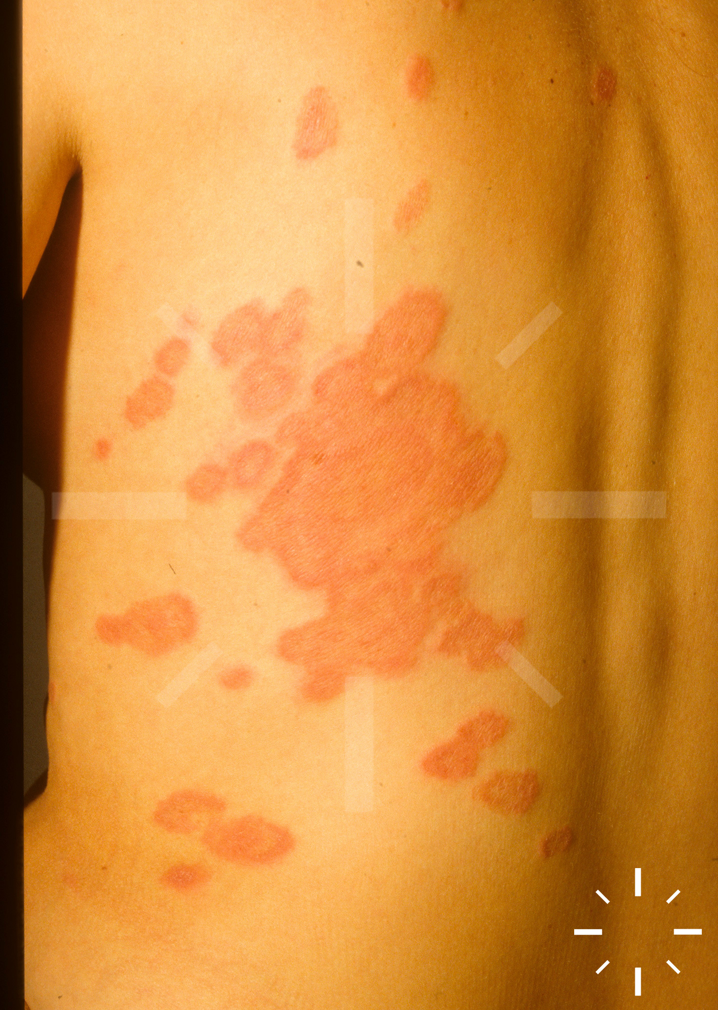
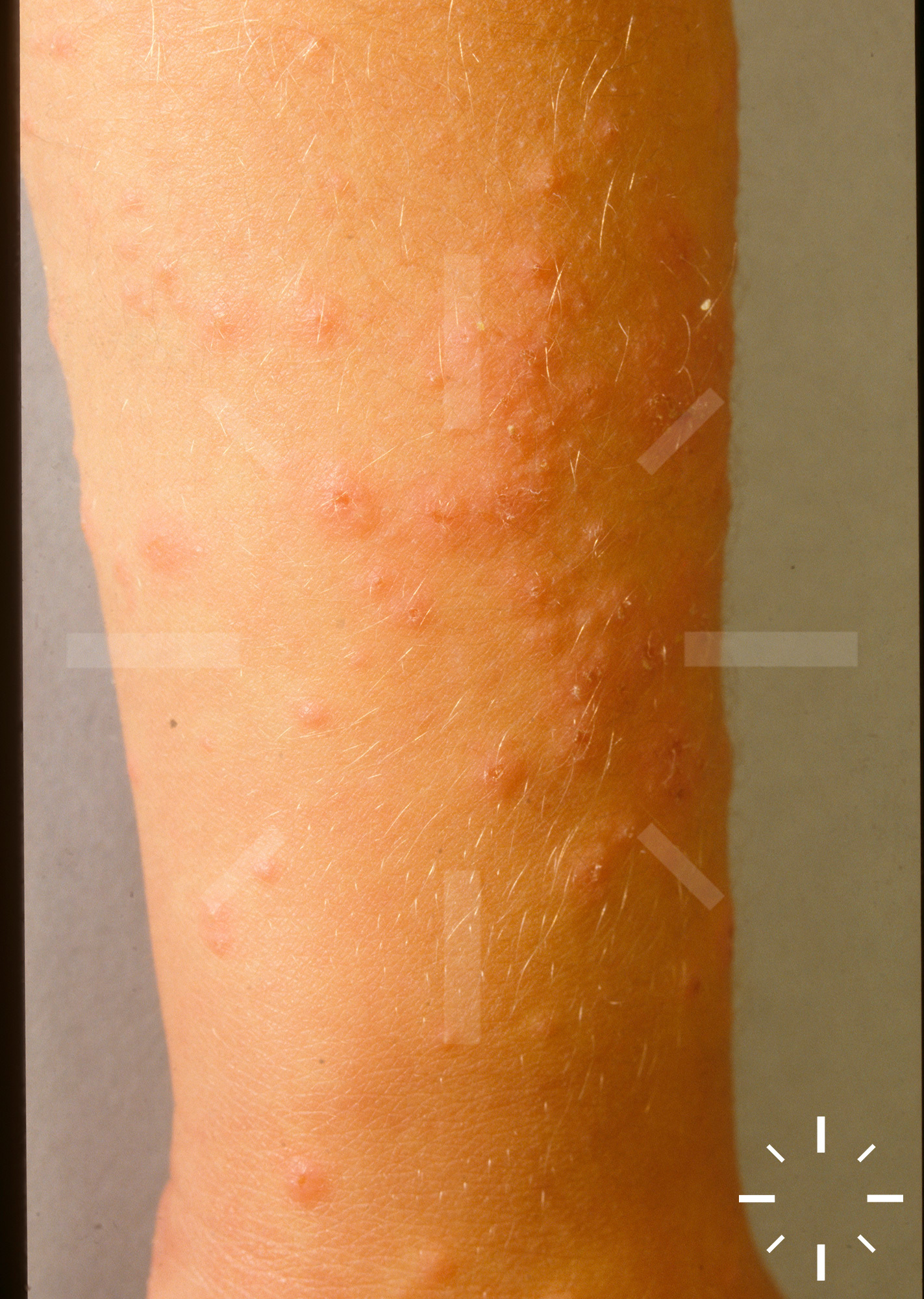
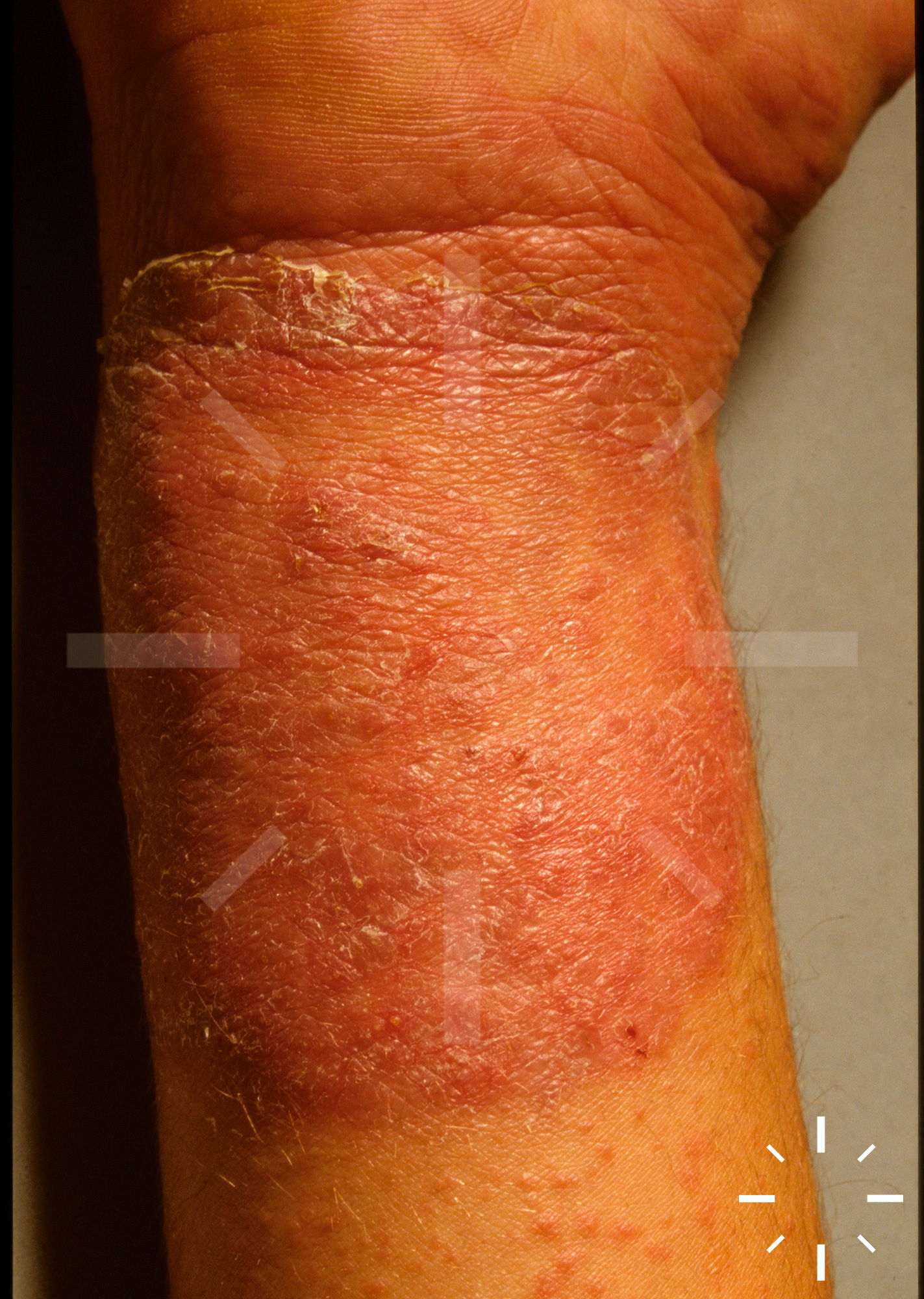
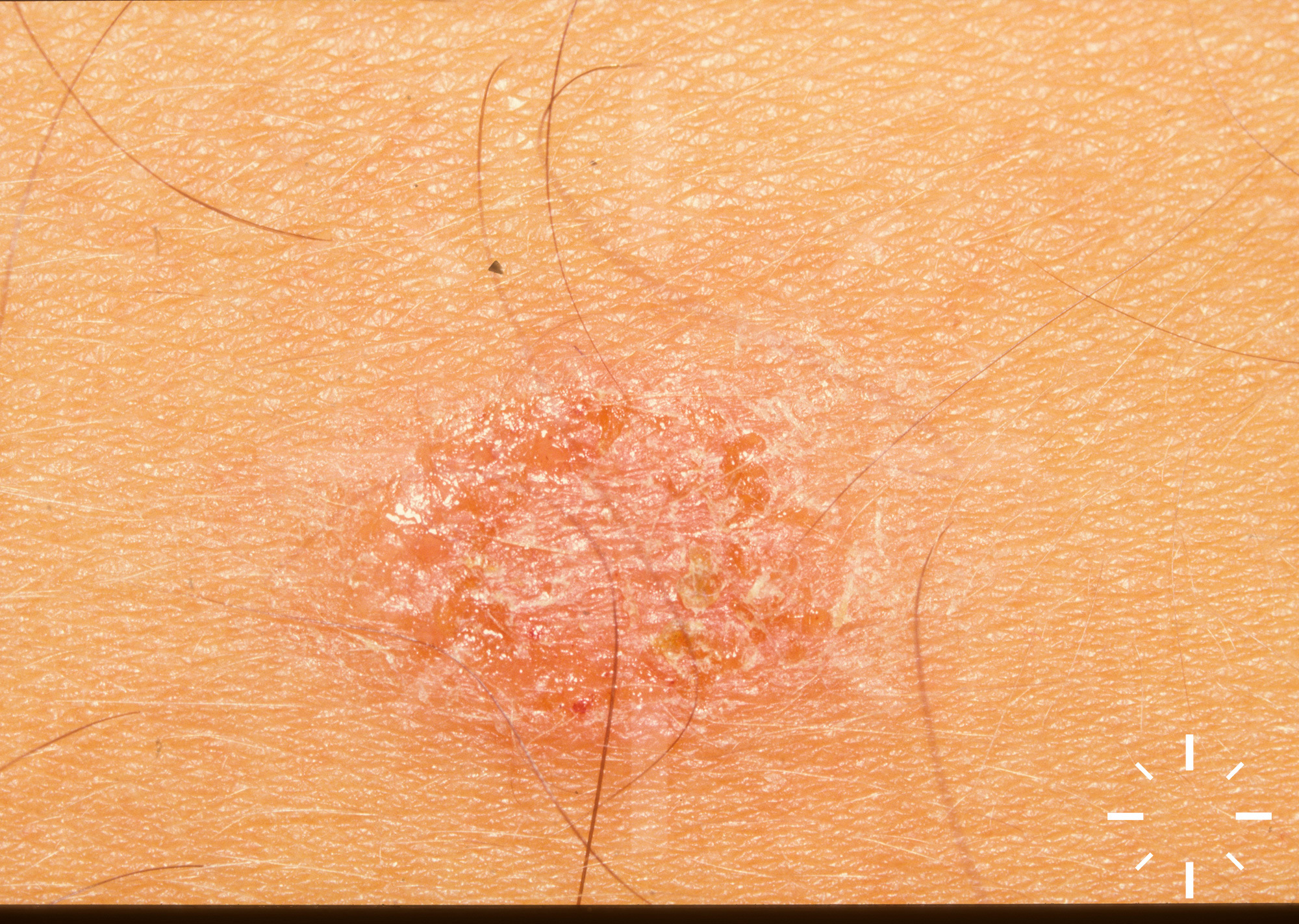
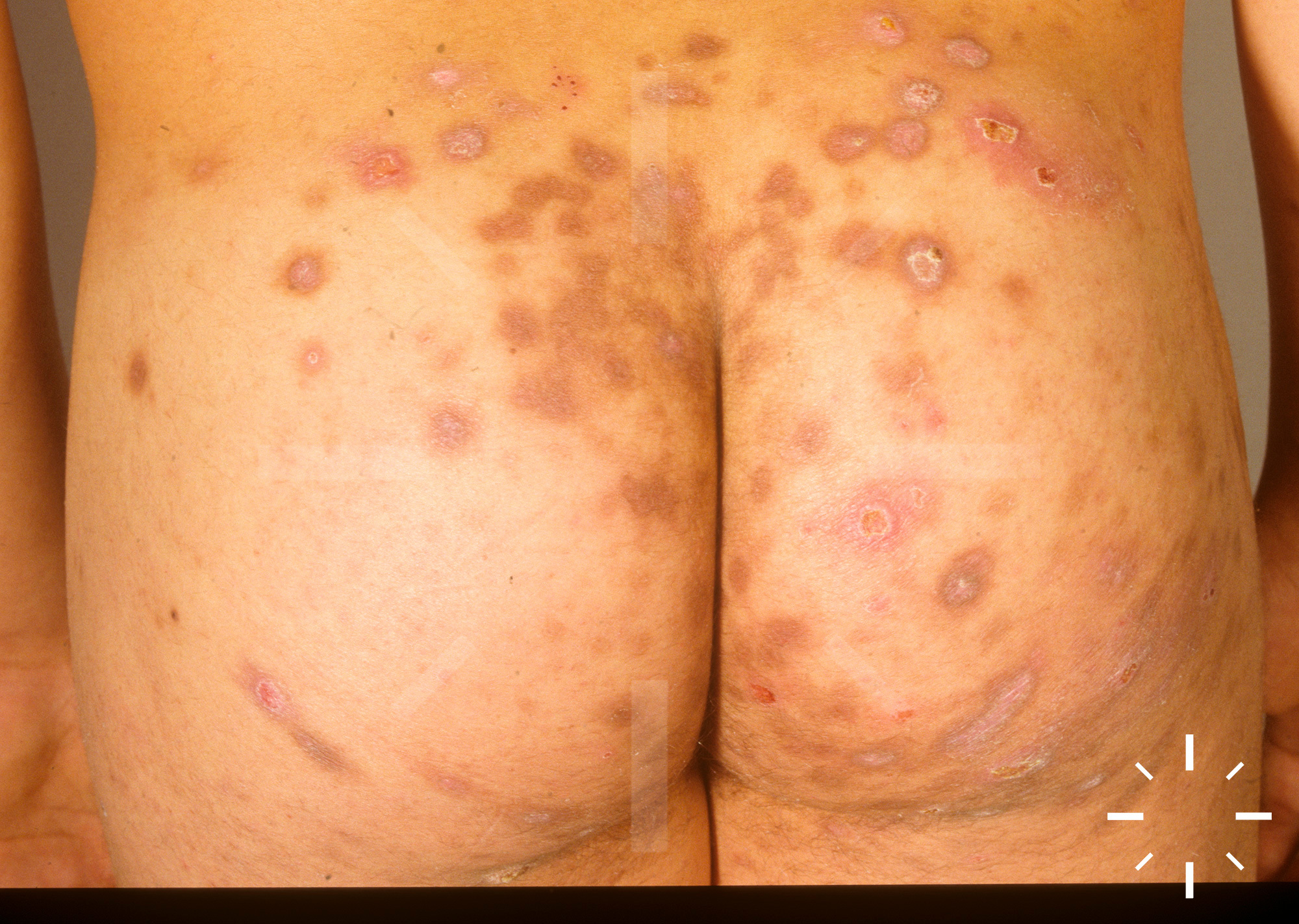
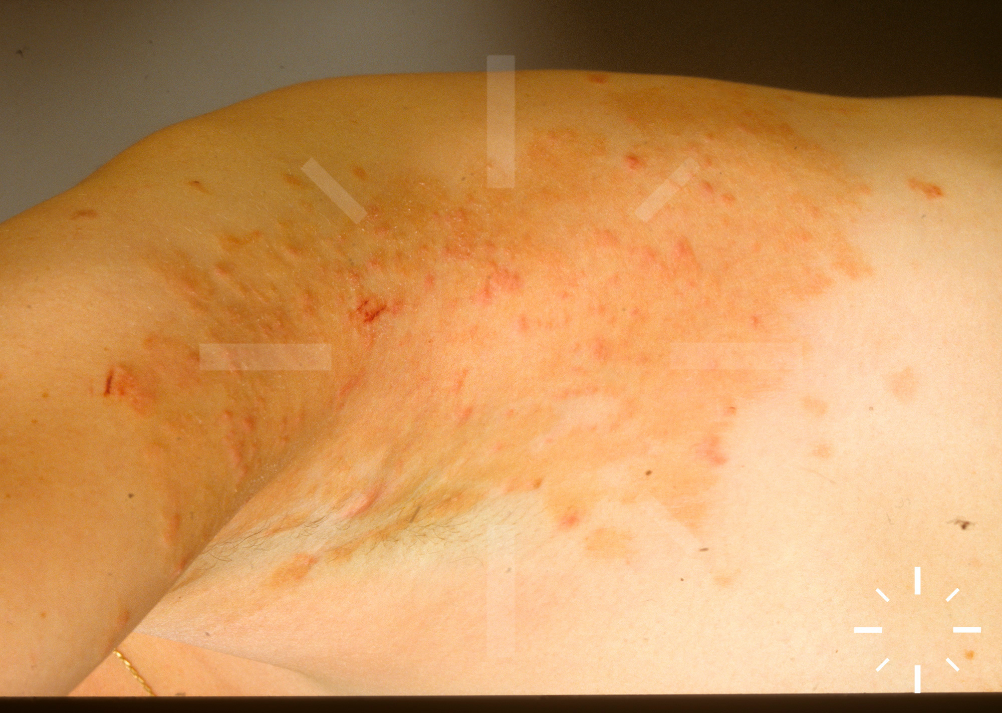
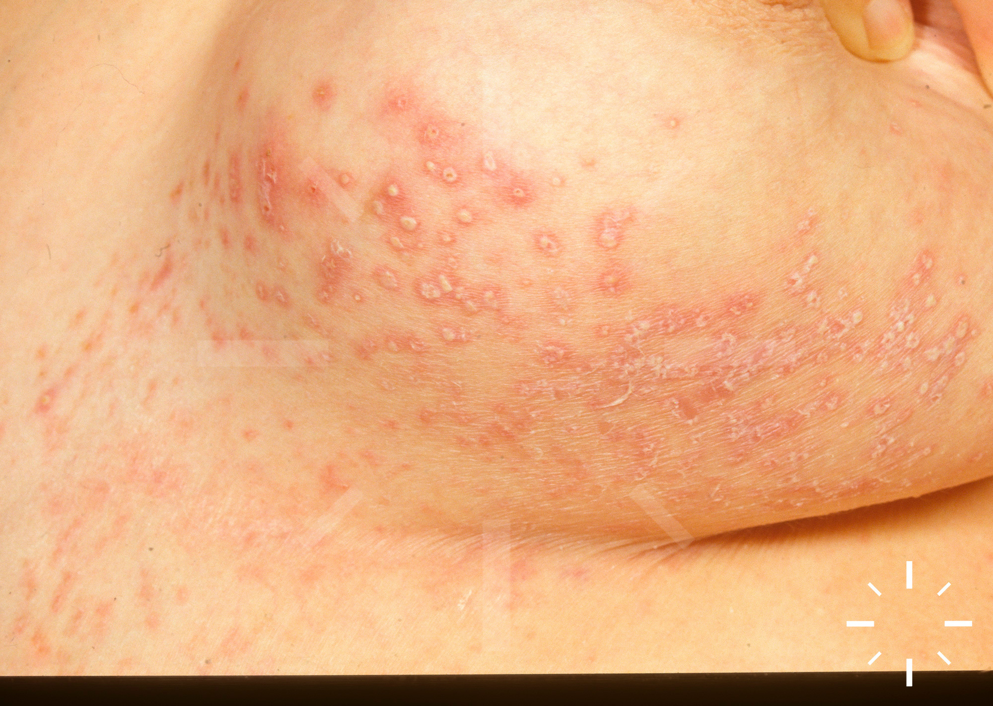
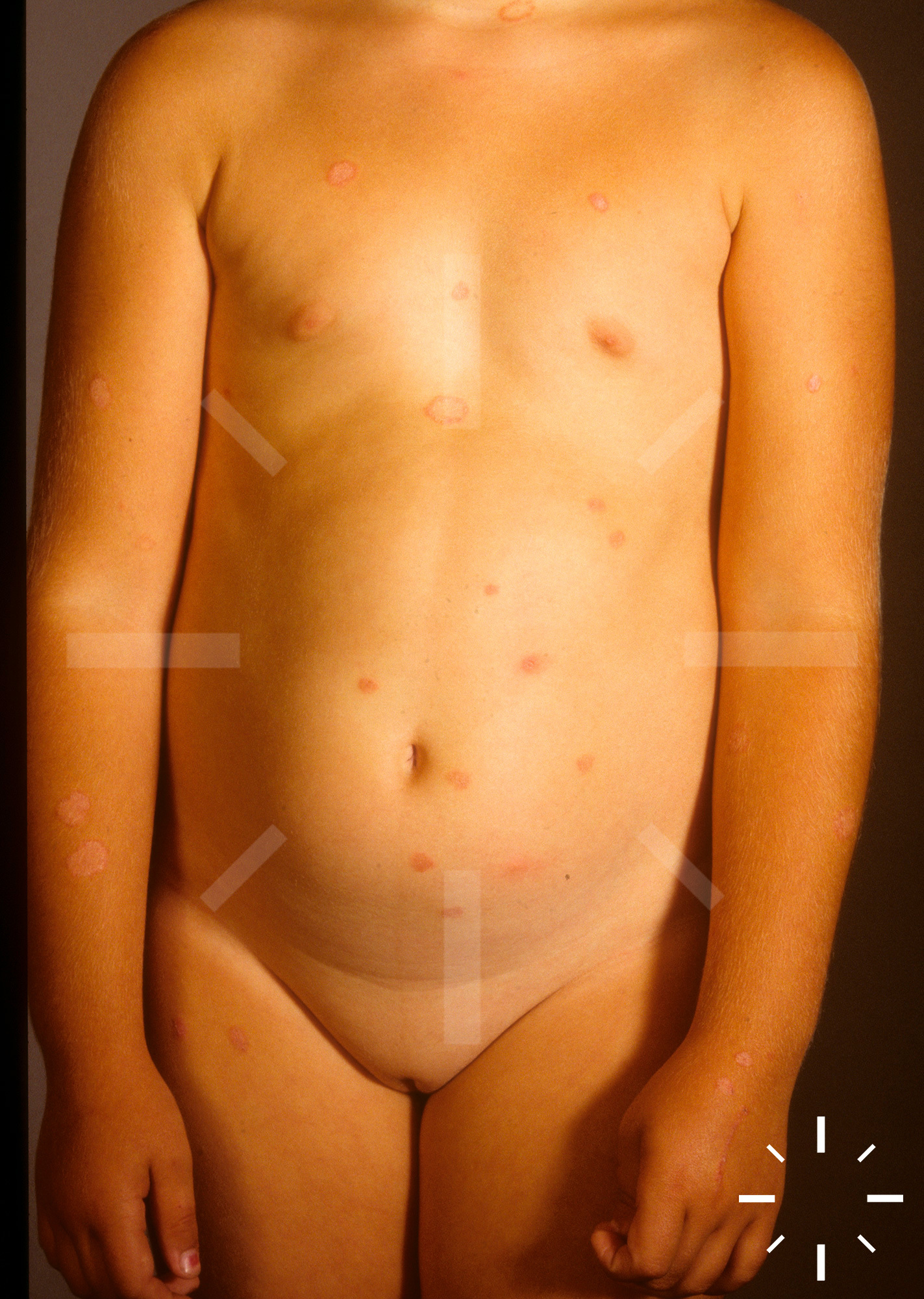
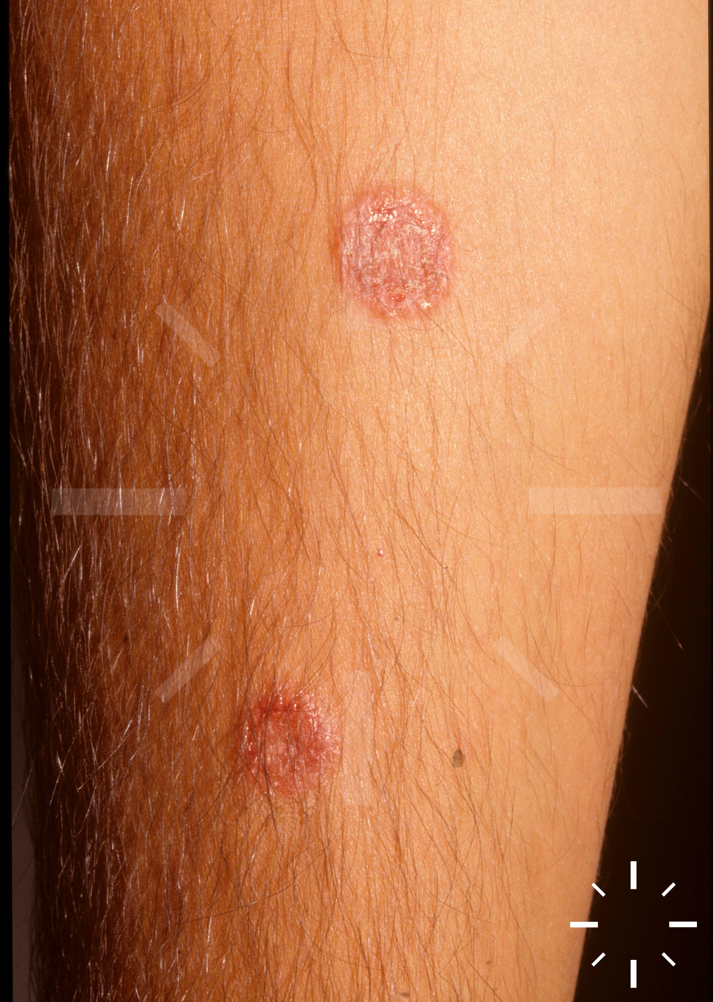
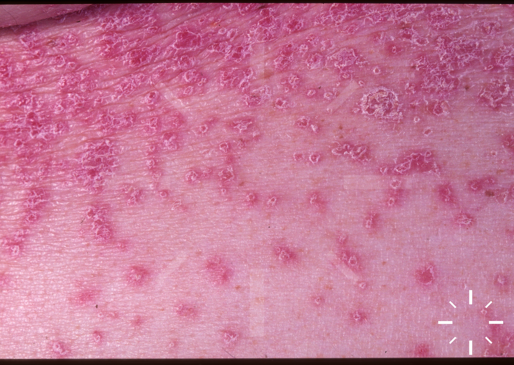
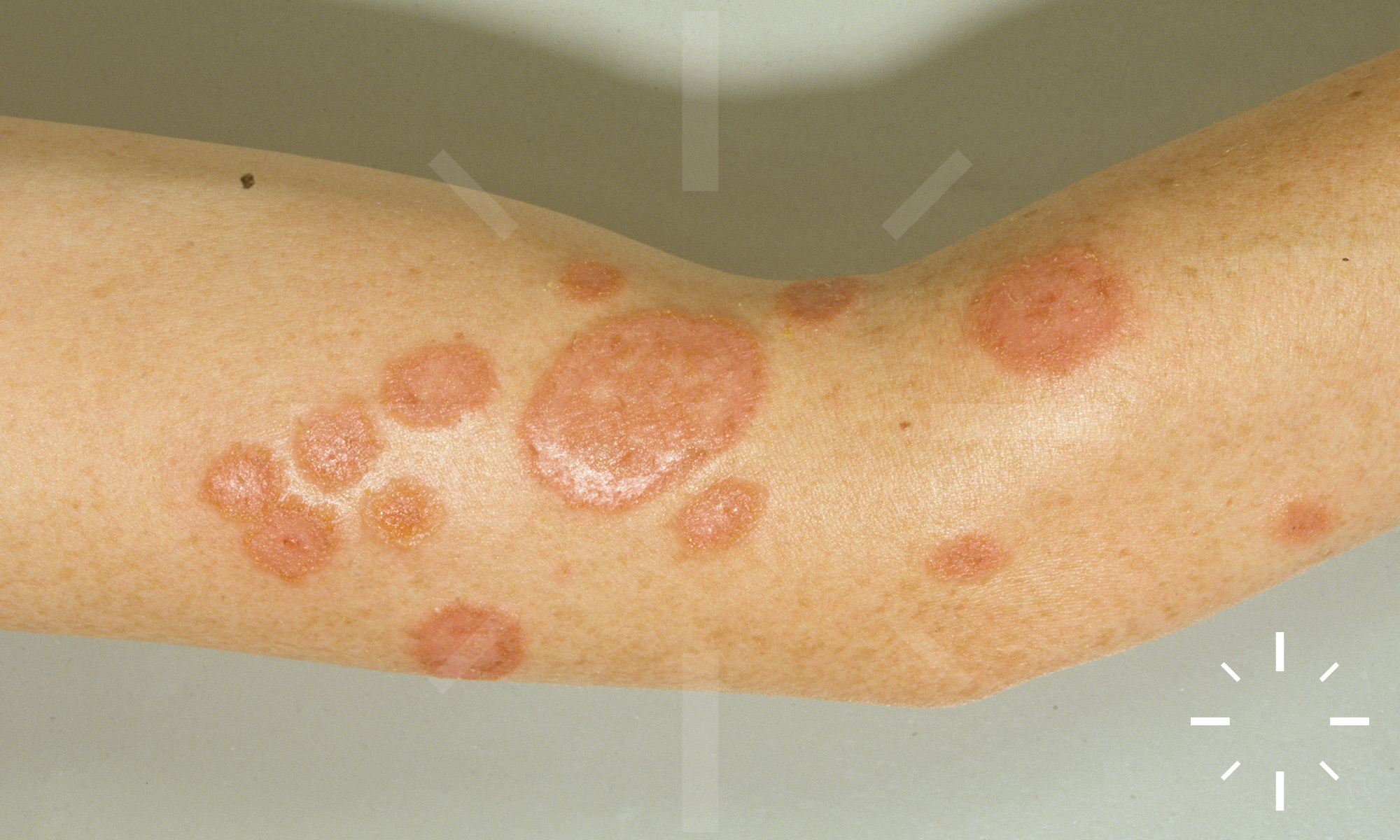
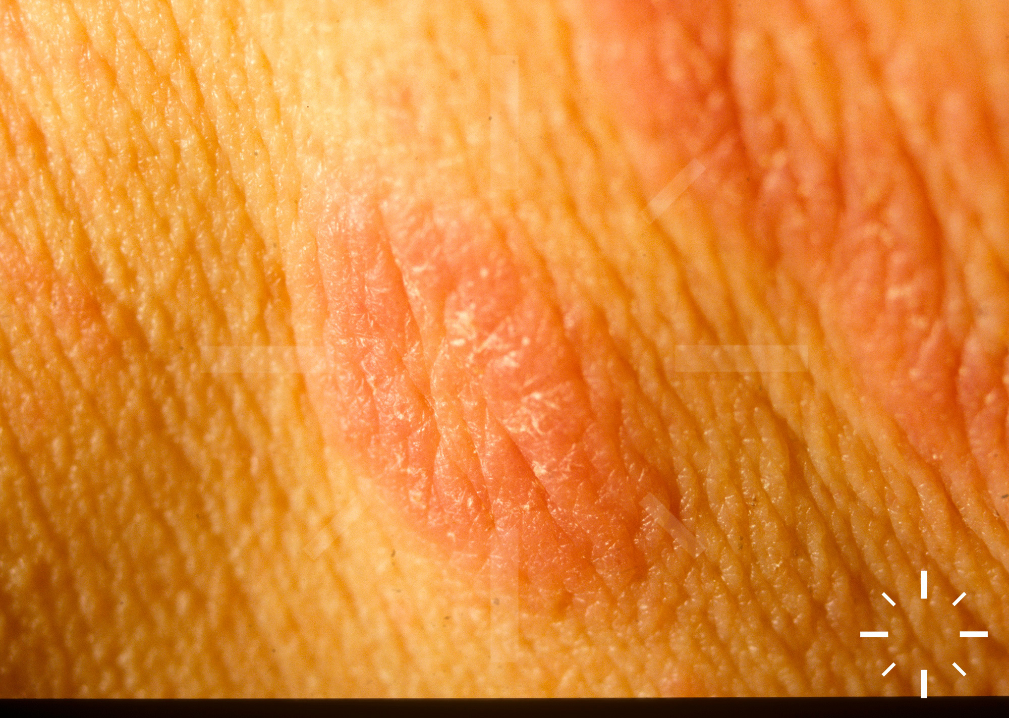
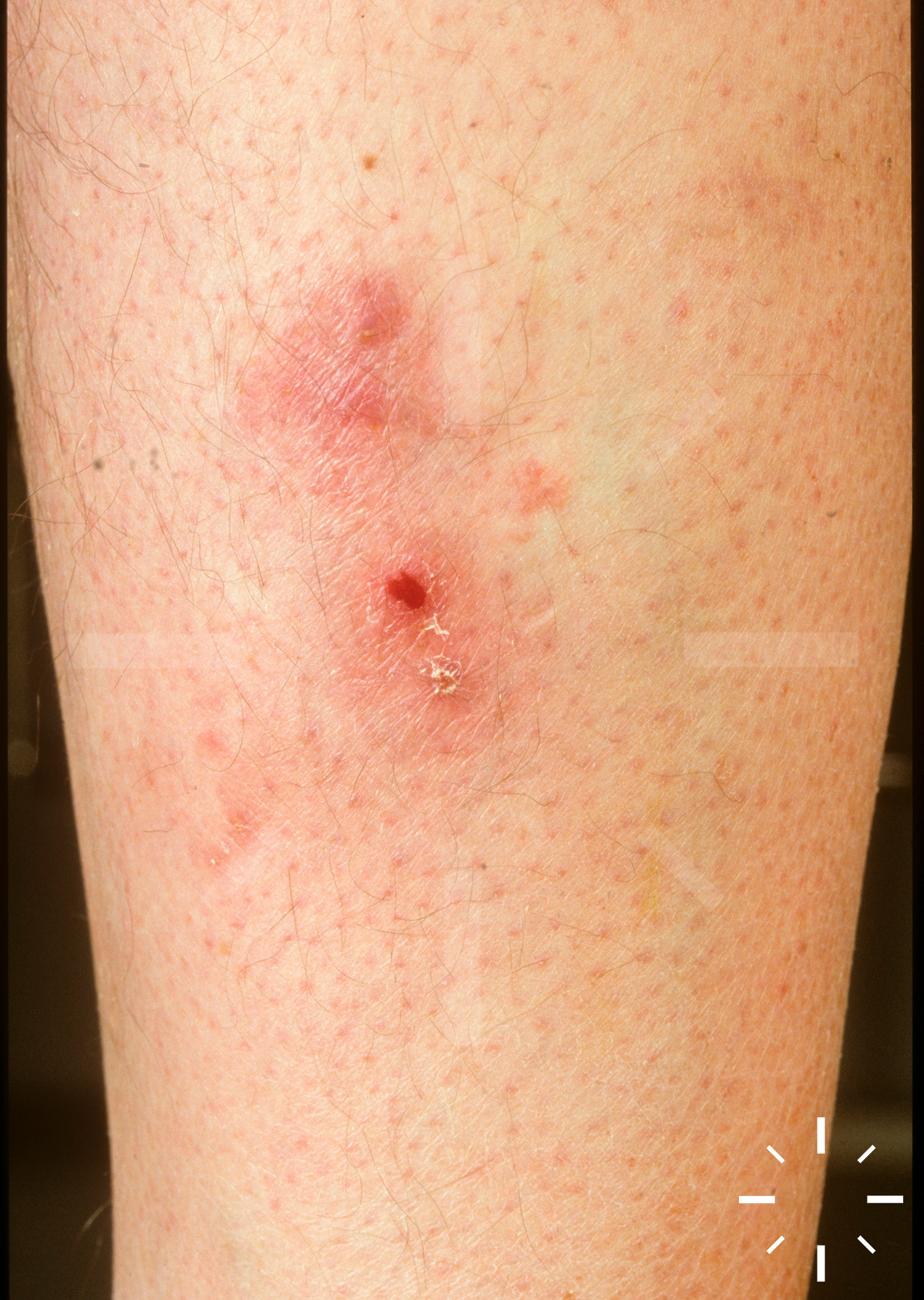
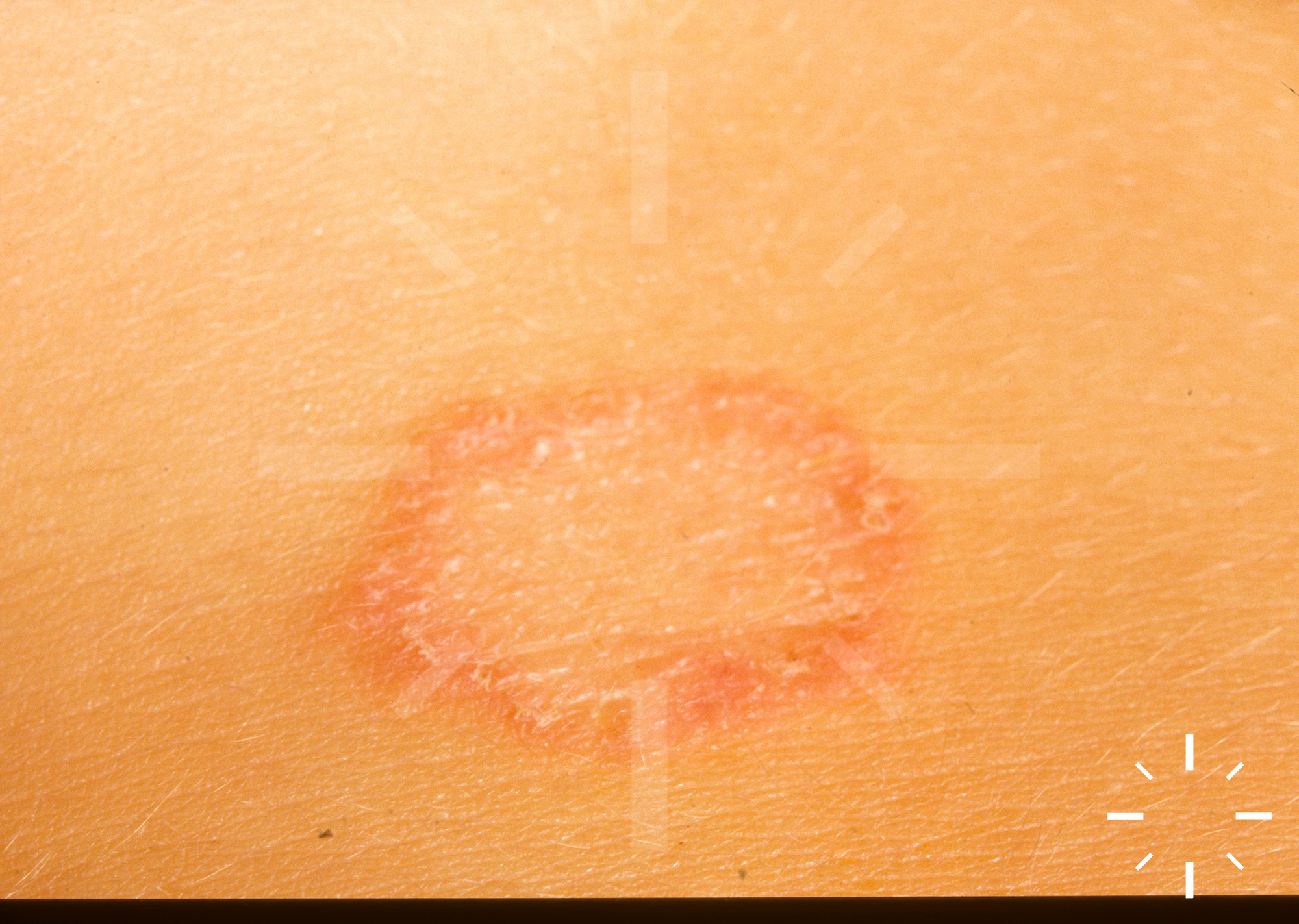
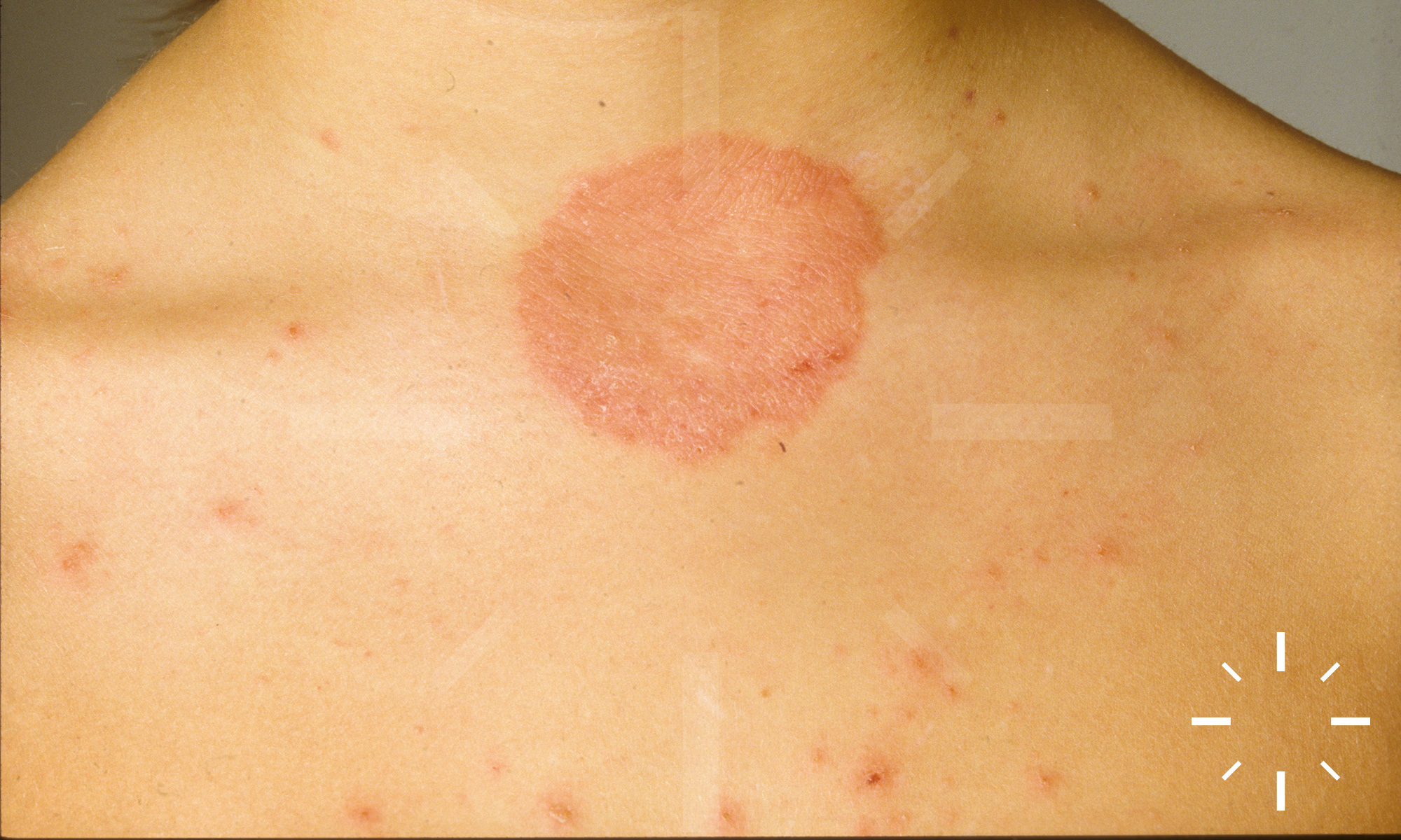
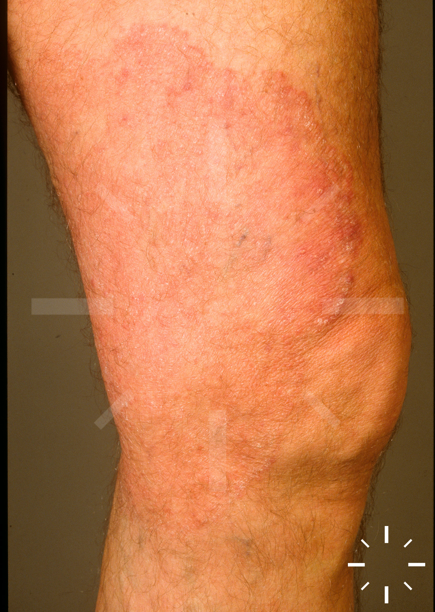
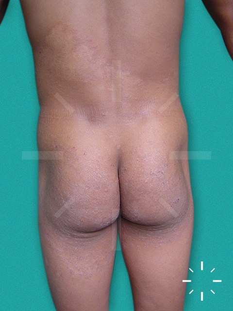
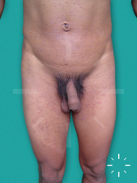
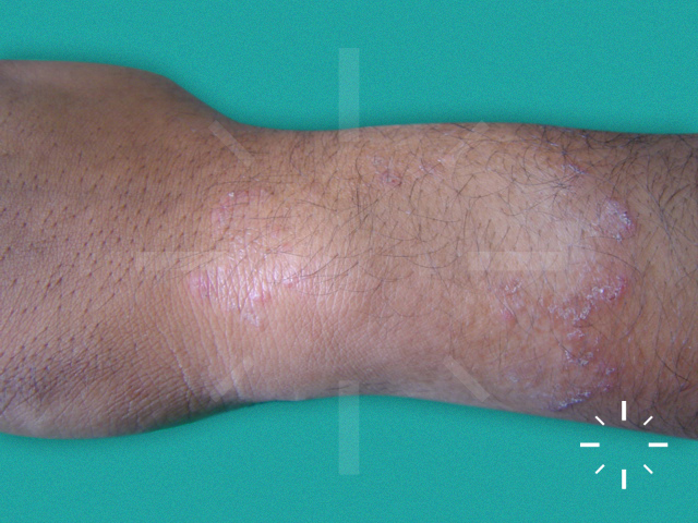
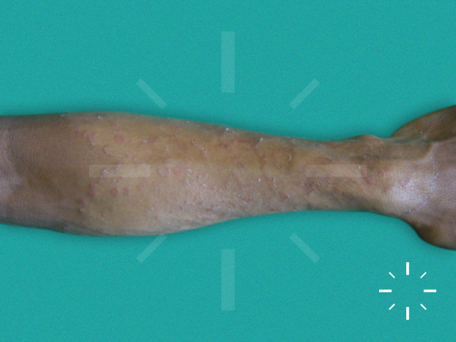
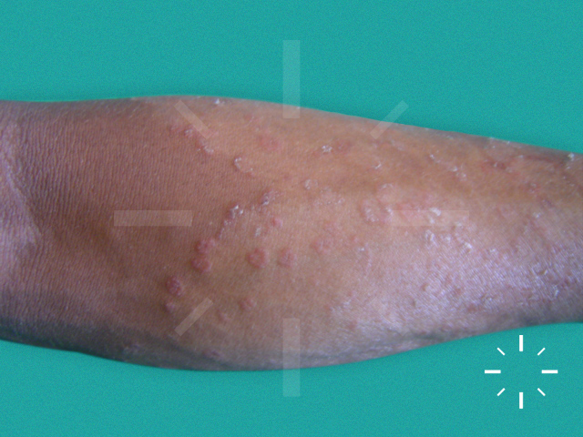
Tinea corporis (mycosis)

Last Updated: 2025-02-11

Author(s): Anzengruber F.

ICD11: 1F28.Y

Trichophytia corporis superficialis.

Superficial infection with dermatophytes.

  • Children > adults
  • Immunosuppression is a predisposing factor.

  • Pathogen:
    • T. rubrum, T. mentagrophytes, Epidermophyton floccosum.
    • Tinea corporis gladiatores is usually caused by T. tonsurans.
  • Transmission often occurs through cats, hamsters or other pets.

  • Initially follicularly bound pustules, which progress in size over time and become erythematous, sharply defined, bordered, pruritic, scaly plaques and asbestos-forming nodules. If they are very widespread, they form map-like figures.

  • Necrosis can also occur, particularly in immunocompromised patients.

  • Chronic tinea corporis is often persistent in body folds.

  • Majocchi granuloma --> Tinea corporis involving the hair follicles and producing pustules and nodules. 

  • Increasingly common: Tinea imbricata, comes from Asia and produces small brownish-red rings. 

  • Animal contact
  • Clinic
  • Detection of pathogen (collection of dandruff: then native preparation and culture)
  • Biopsy with PAS staining
  • Wood light (discrete fluorescence)

  • Possible on the entire integument

  • Spontaneous healing possible
  • Often, especially without therapy, highly chronic course.

  1. Voravutinon V. Oral treatment of tinea corporis and tinea cruris with terbinafine and griseofulvin: a randomised double blind comparative study. J Med Assoc Thai 1993;76:388-93.
  2. Farag A, Taha M, Halim S. One-week therapy with oral terbinafine in cases of tinea cruris/corporis. Br J Dermatol 1994;131:684-6.