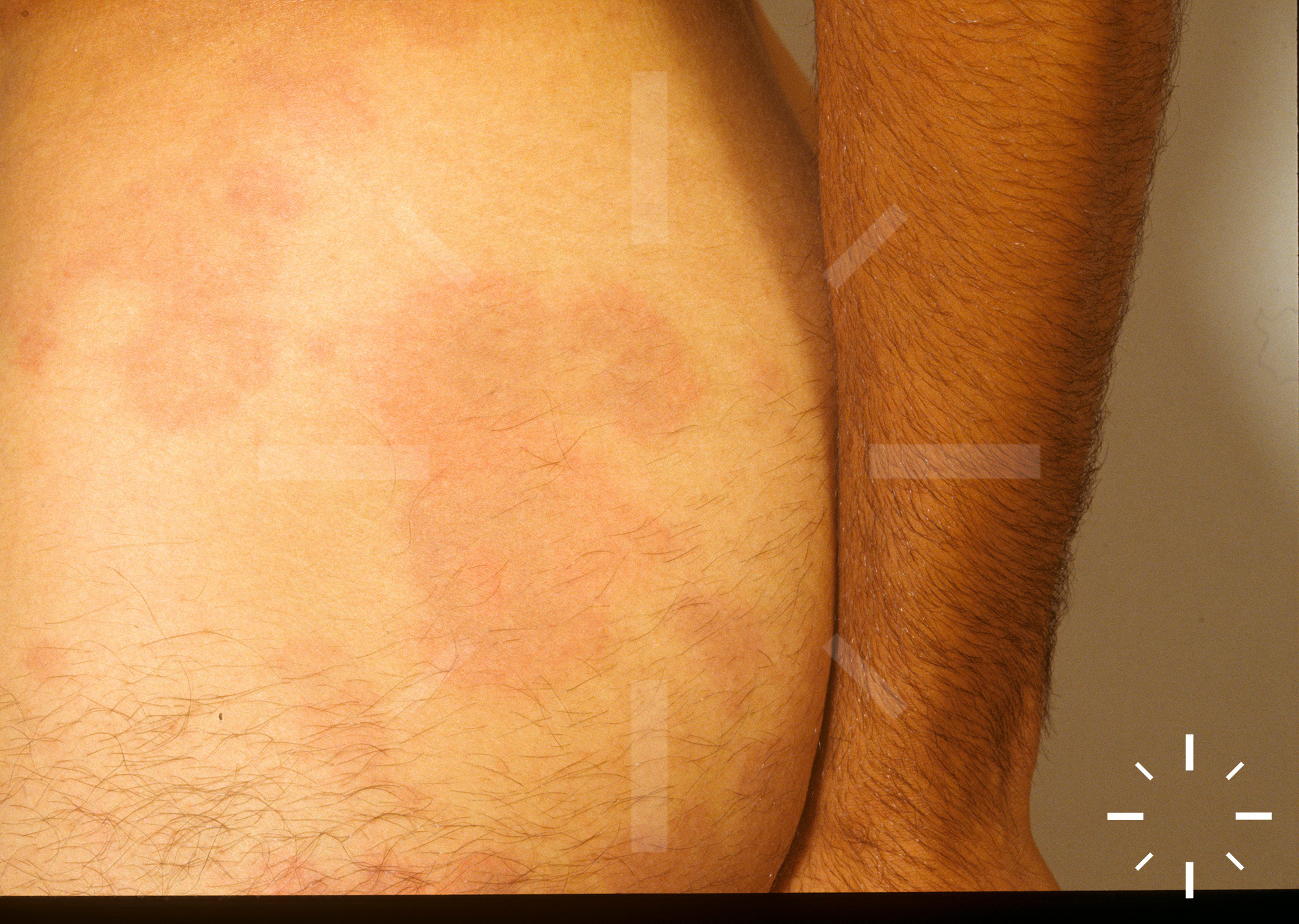
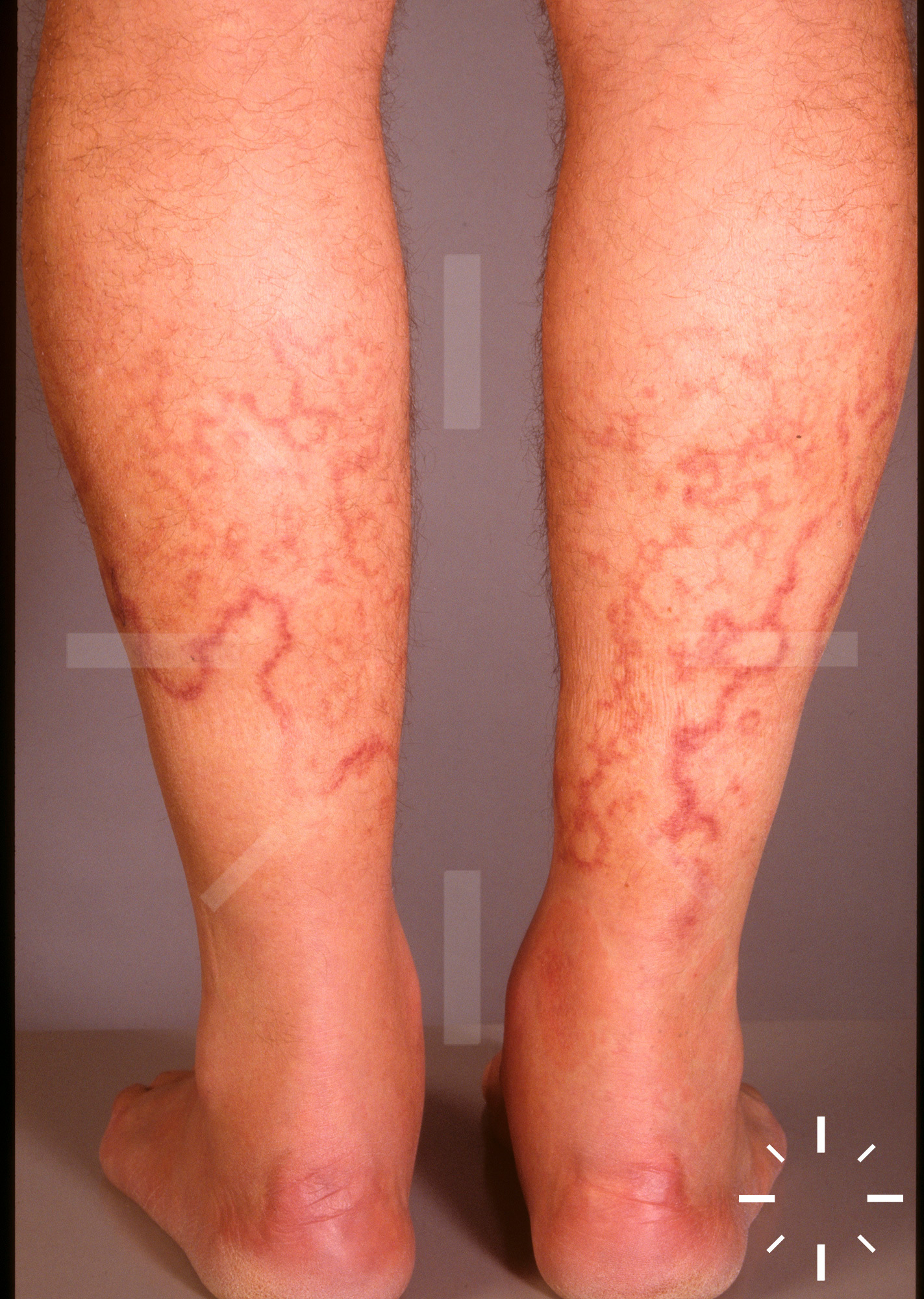
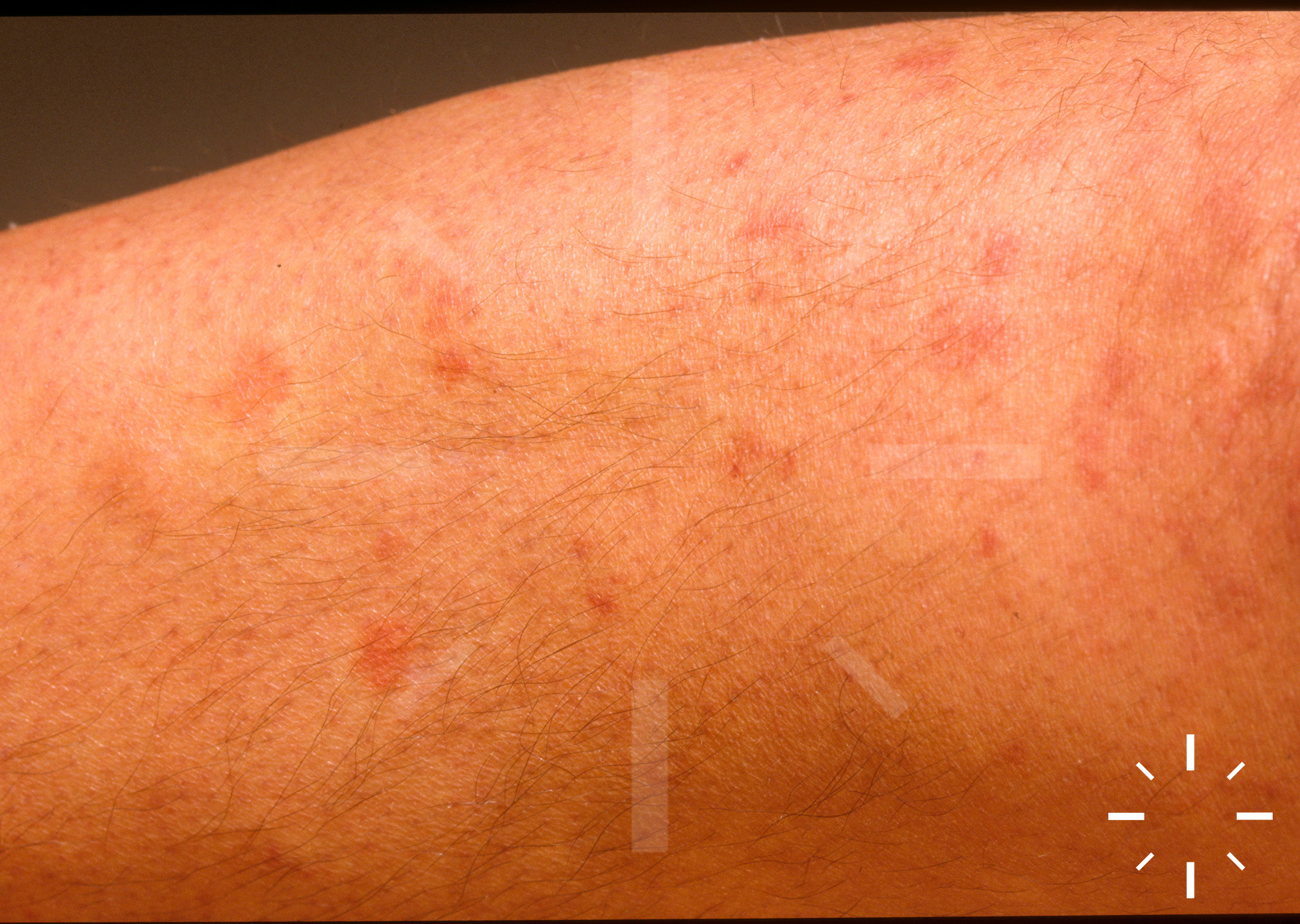
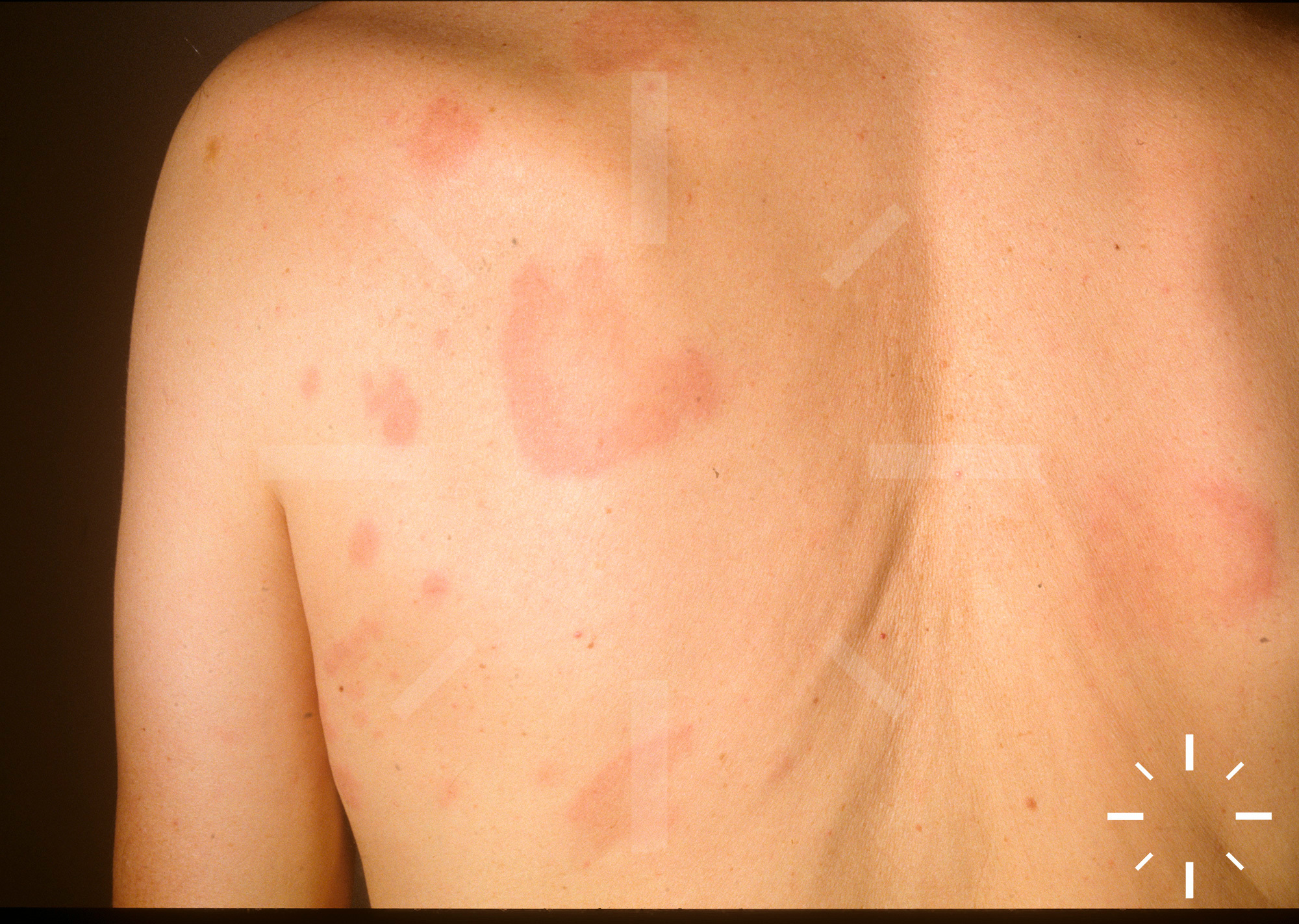
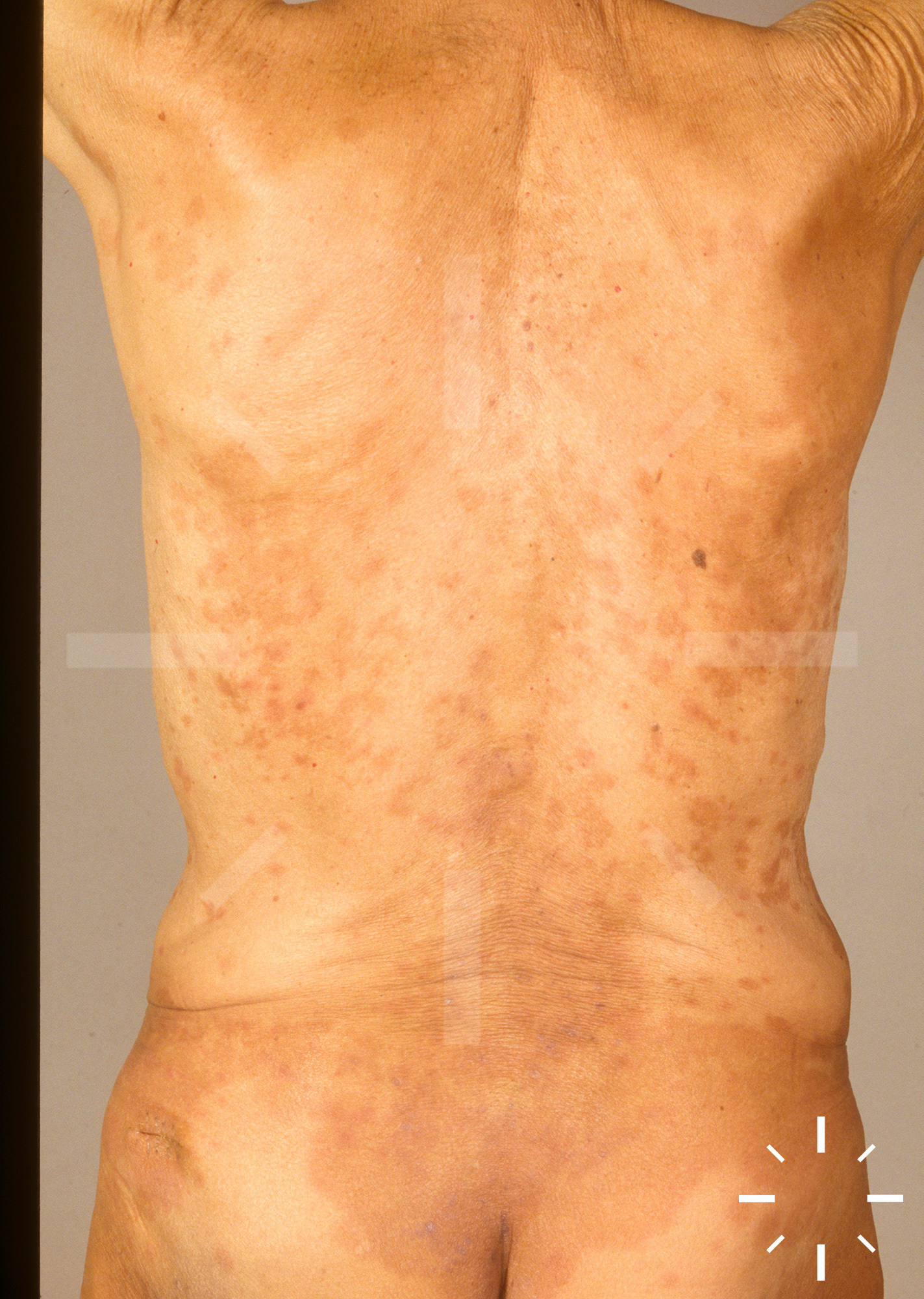
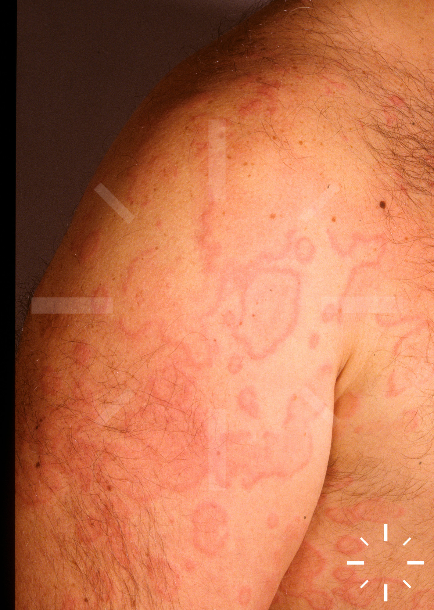
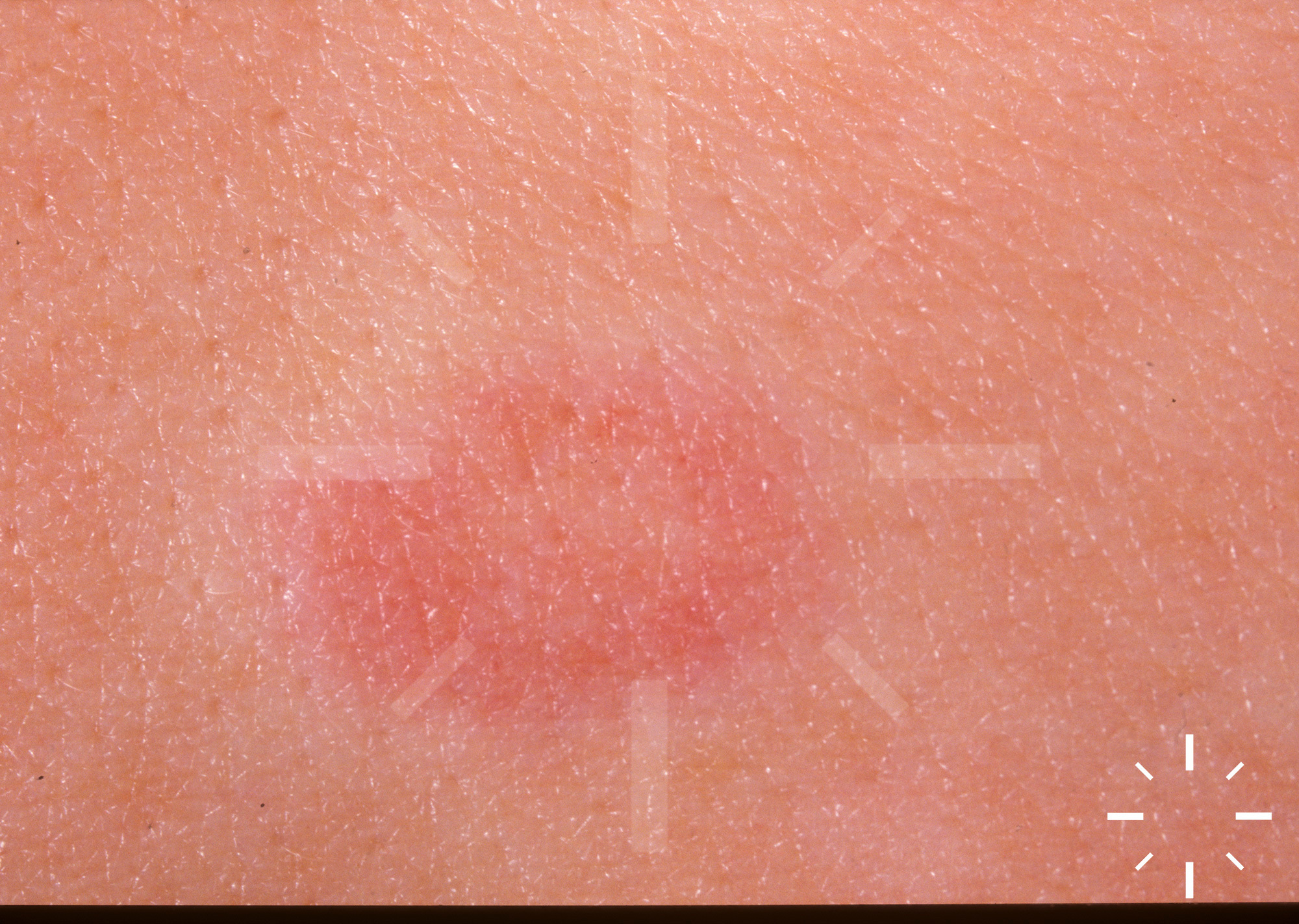
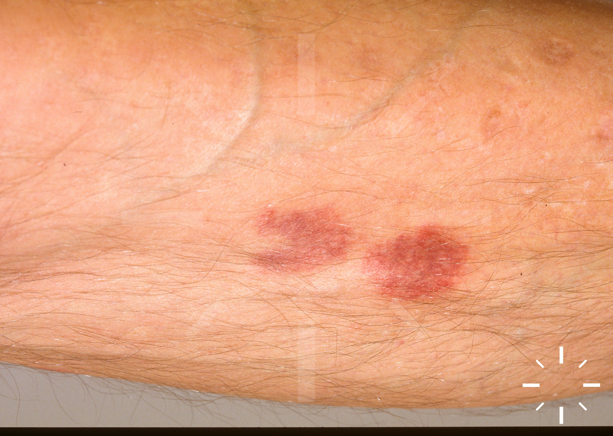
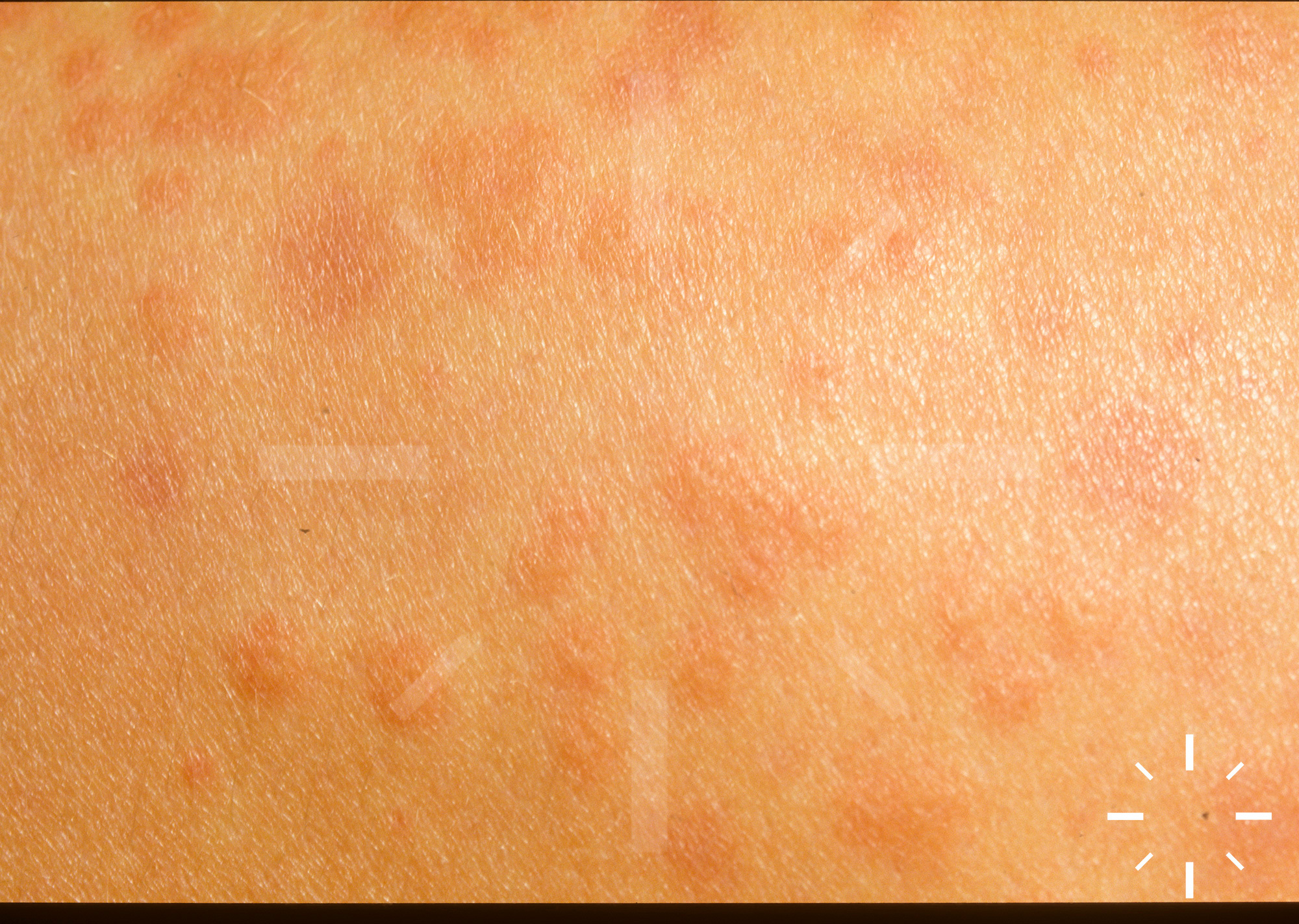
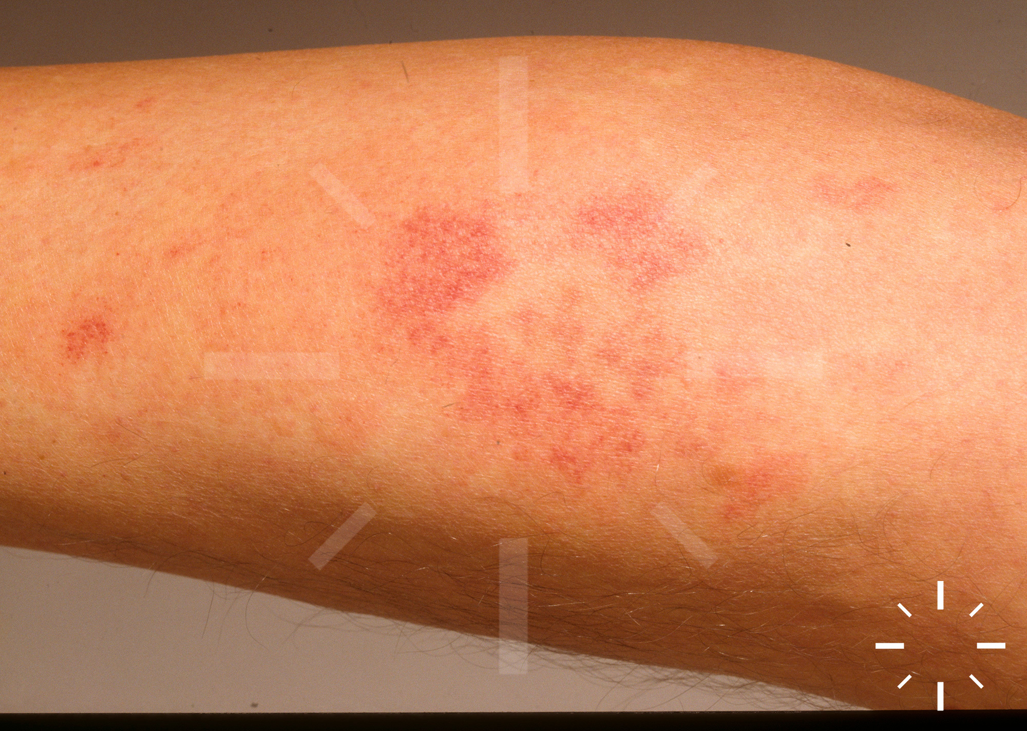
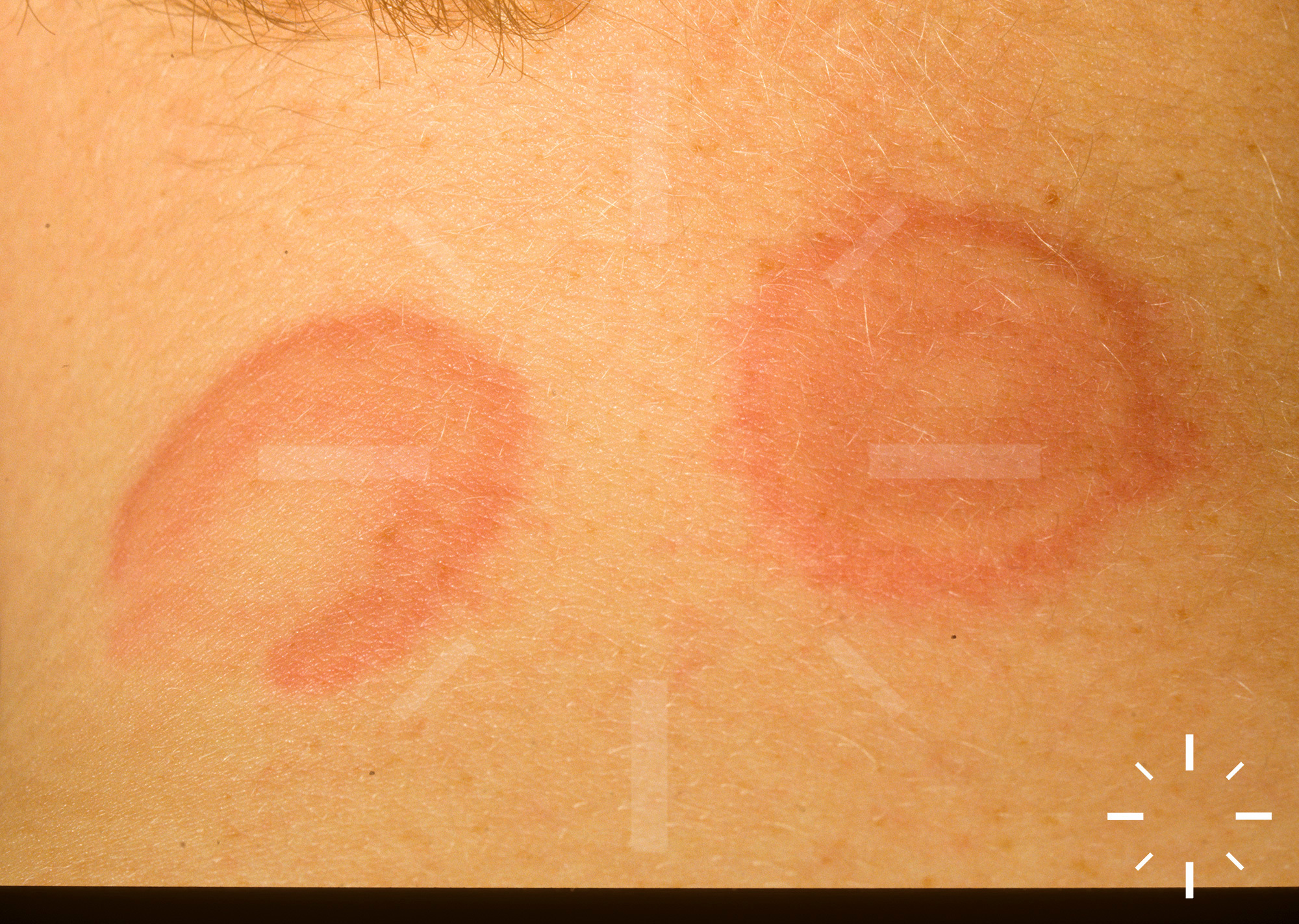
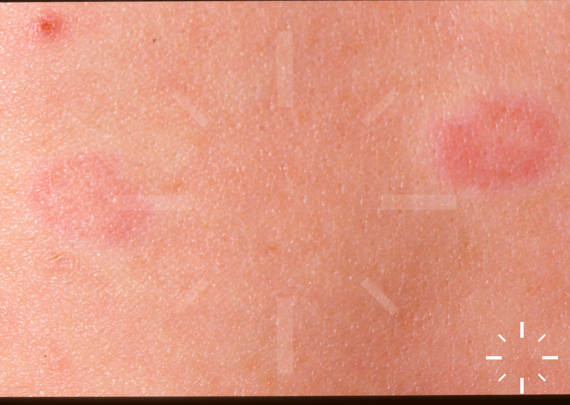
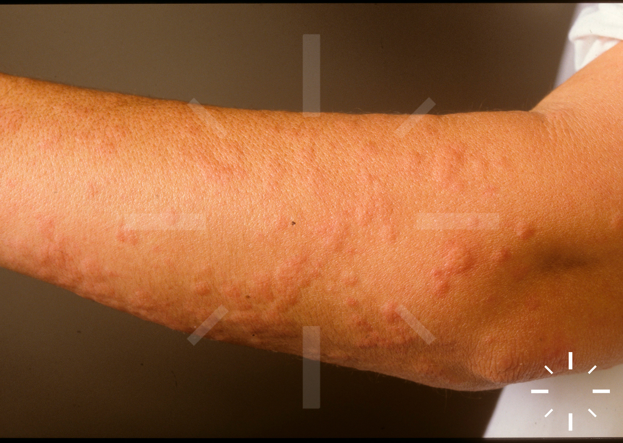
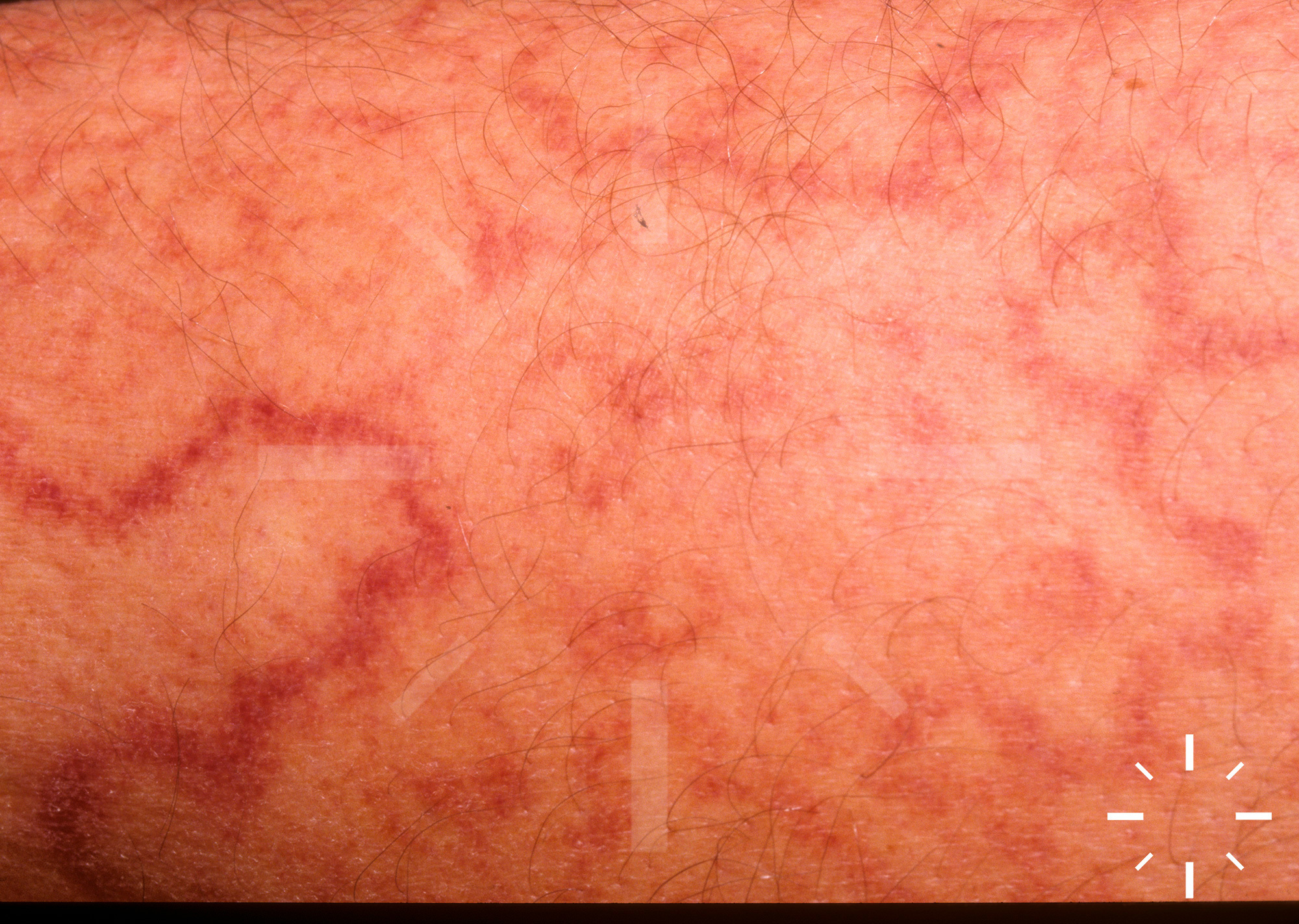
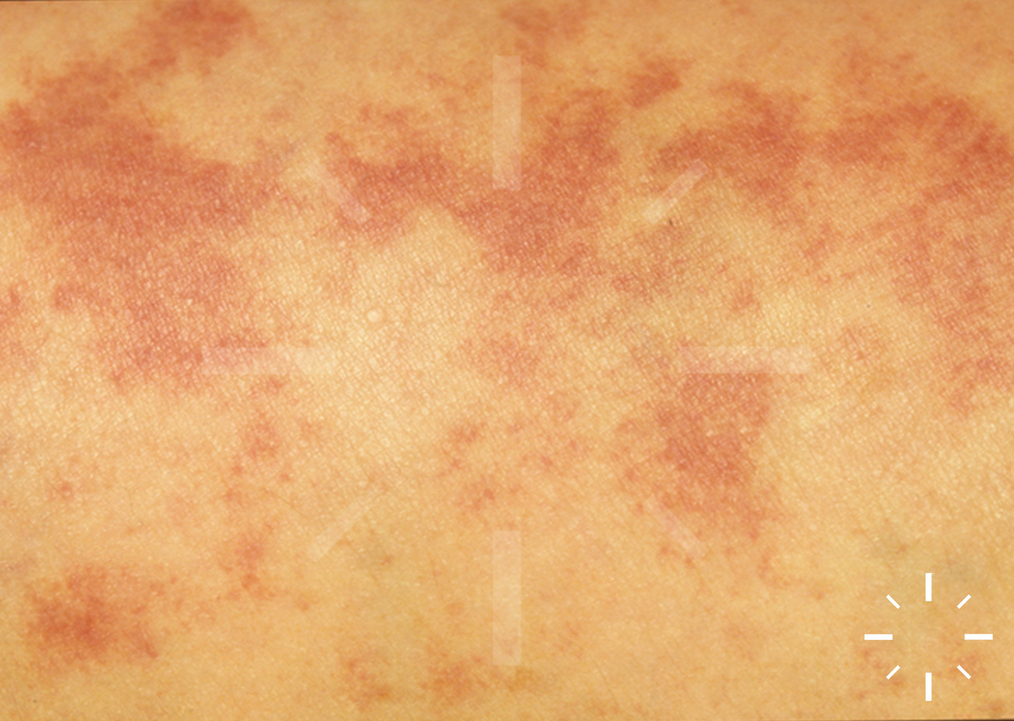
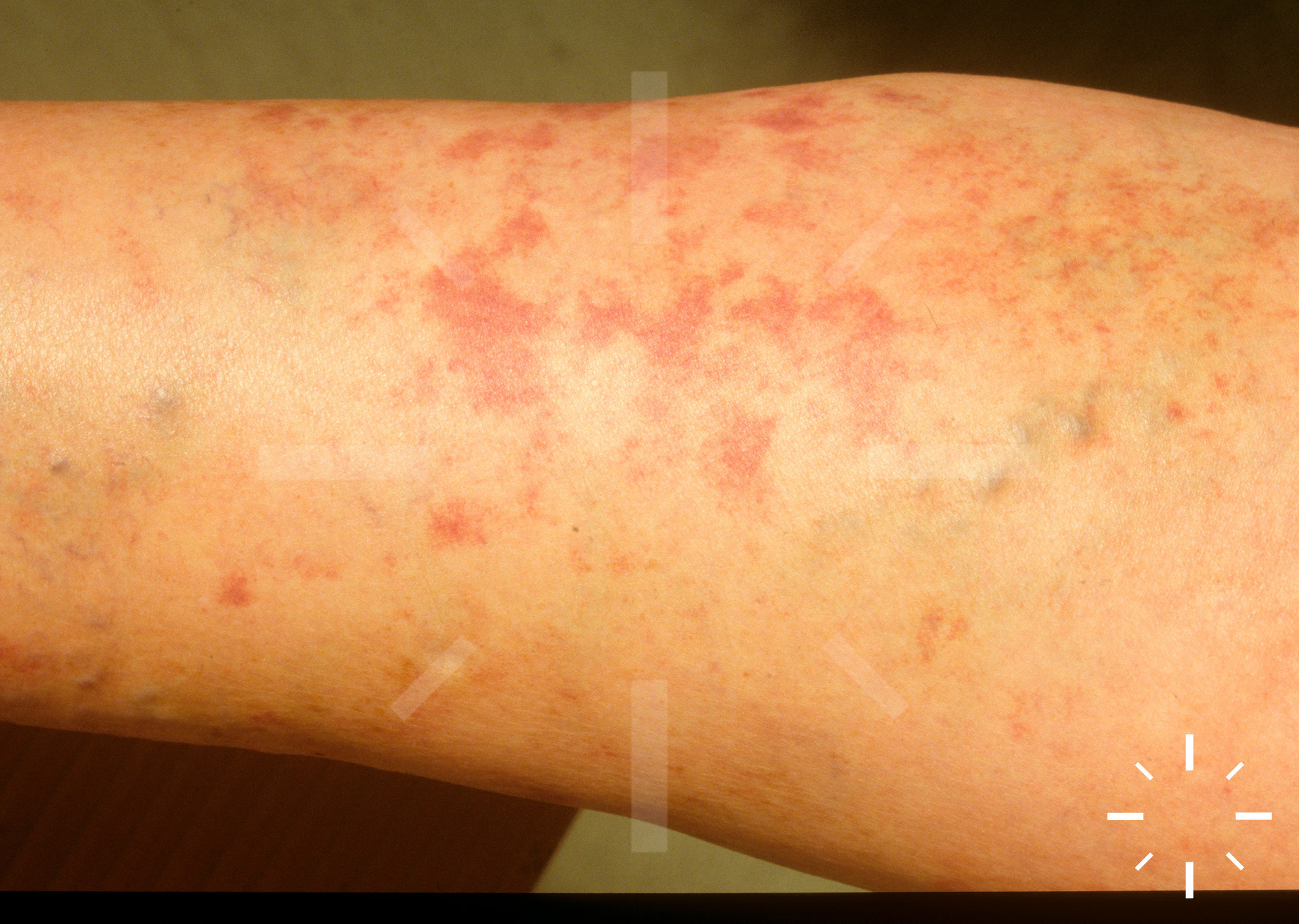
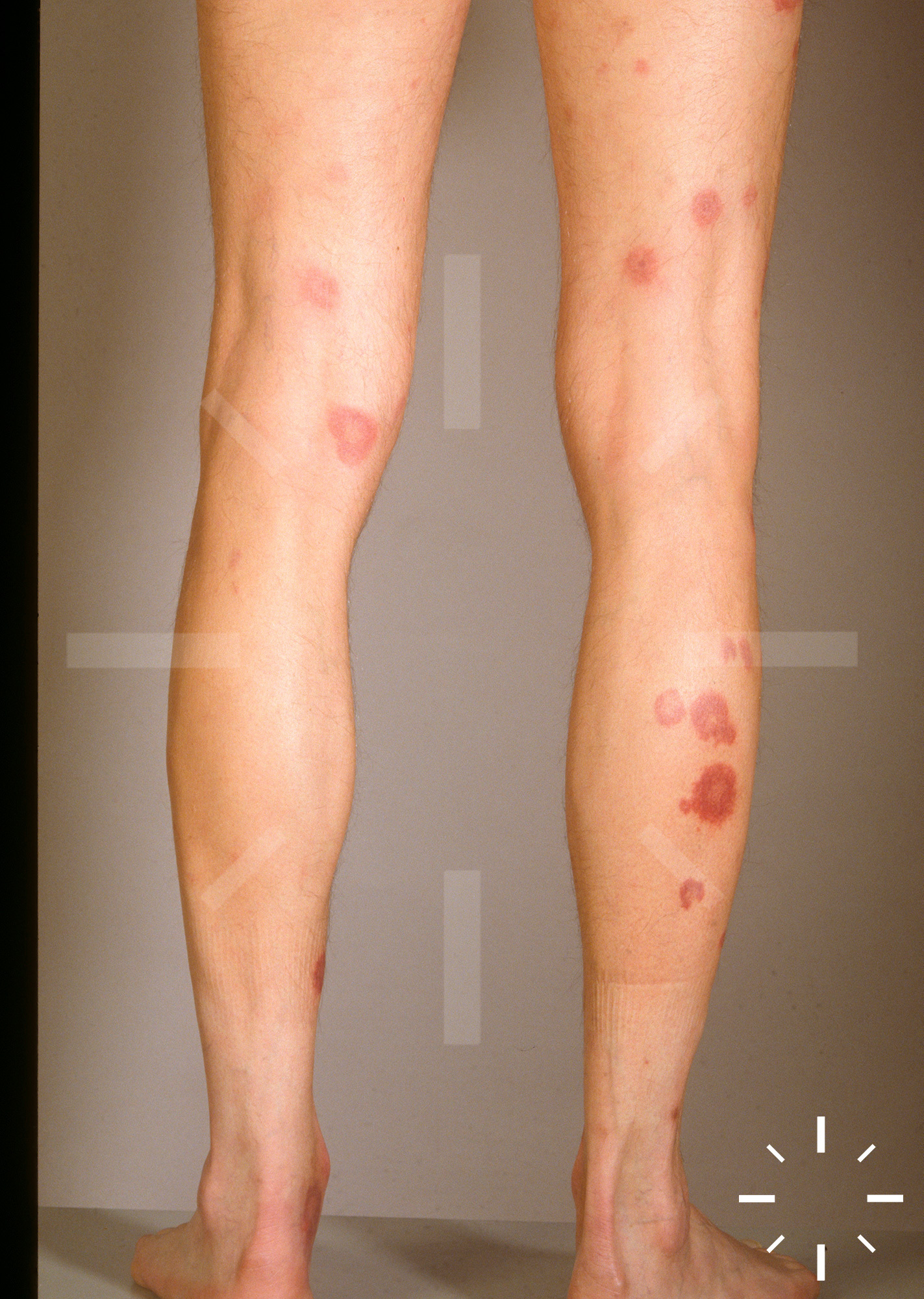
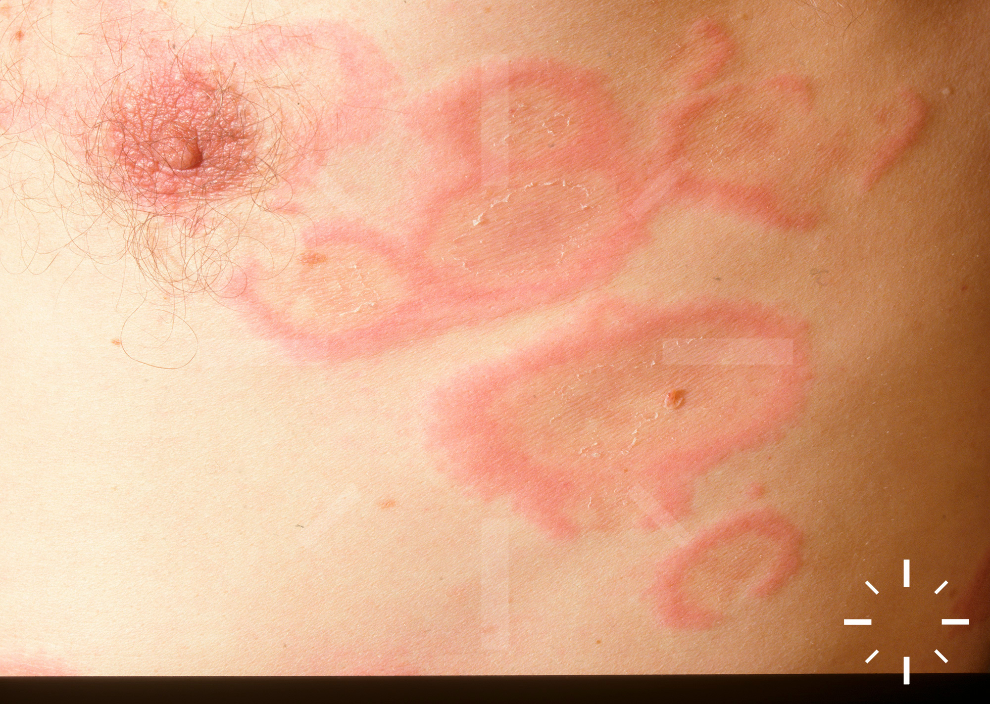
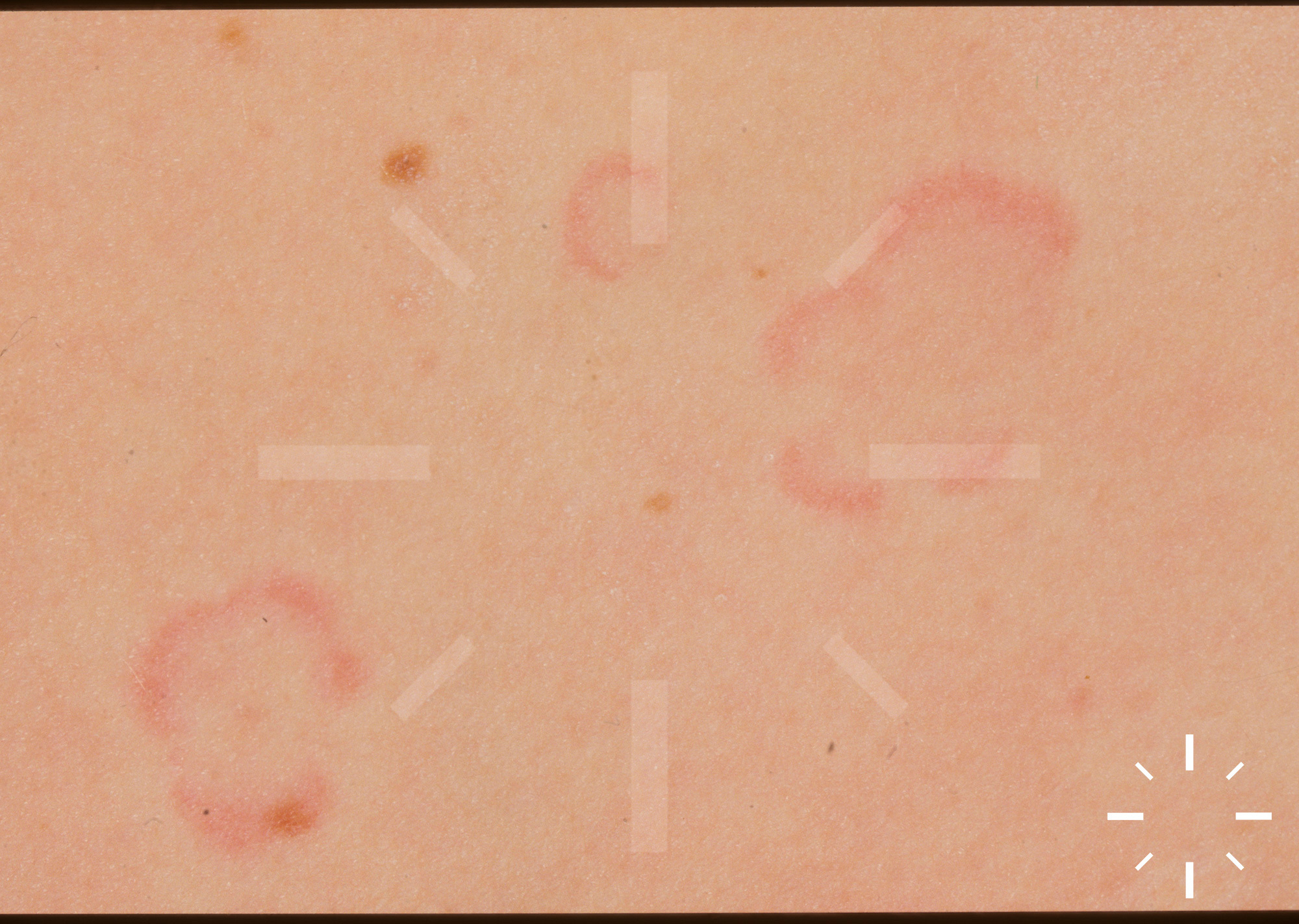
Urticarial vasculitis
Last Updated: 2025-02-11
Author(s): Anzengruber F., Navarini A.
ICD11: EF40.10

1/20